


Abstract
The histamine H1 receptor (H1R)-mediated signaling cascade is inhibited by phorbol ester-induced protein kinase C (PKC) activation. Cloning studies of the H1Rs have shown that several potential PKC phosphorylation sites are located in the third intracellular loop of H1R. To elucidate the molecular mechanism of PKC-mediated desensitization, we identified amino acid residues that are involved in the desensitization of the H1R. Two amino acid residues (Ser396, Ser398) were determined to be PKC phosphorylation sites by in vitro phosphorylation studies using a series of synthetic peptides. Treatment with phorbol ester decreased histamine-induced accumulation of inositol phosphates in Chinese hamster ovary cells expressing the H1R with a rightward shift in the EC50 value, which implies the uncoupling of the receptor from the G protein. Site-directed mutagenesis studies showed that substitution of alanine for Ser398 but not for Ser396 markedly attenuated the effect of phorbol ester, which suggests that the Ser398 residue was primarily involved in PKC-mediated desensitization.
Histamine receptors are classified into three subtypes (Haaksma et al., 1990;Hill, 1990), based on pharmacological studies and gene cloning. The histamine H1 receptor (H1R) is mainly involved in immune hypersensitivity in peripheral tissues and neurotransmission in the central nervous system (Fukui and Yanai, 1993). Thus, H1R antagonists are used in the treatment of allergic disorders, insomnia, anorexia, and motion sickness. The H1R, a G protein-coupled receptor, associates with phospholipase C (PLC), and stimulation of the receptor leads to the formation of two second messengers, inositol 1,4,5-trisphosphate (IP3) and 1,2-diacylglycerol (Haaksma et al., 1990; Hill, 1990). IP3 induces the release of Ca2+ from intracellular stores, and 1,2-diacylglycerol activates protein kinase C (PKC).
Loss of biological response (desensitization) is induced to protect cells from excess stimulation. Two types of desensitization have been observed in G protein-coupled receptors. Homologous desensitization is agonist-specific, whereas heterologous desensitization is not. Both types of desensitization seem to be mediated by phosphorylation of the receptor, but the phosphorylation site and phosphorylating enzyme are different for each receptor. In the case of the β-adrenergic receptor, homologous desensitization is mediated by phosphorylation of the carboxyl-terminal tail of the receptor by G protein-coupled receptor kinase (GRK) (Bouvier et al., 1988; Lohse et al., 1990), whereas heterologous desensitization is mediated by phosphorylation of the third intracellular loop of the receptor by second messenger-dependent protein kinases such as cAMP-dependent protein kinase (cAPK) or PKC (Yuan et al., 1994). However, the molecular mechanisms of homologous and heterologous desensitization of many other G-protein-coupled receptors remain unclear.
Pretreatment with histamine causes homologous desensitization of the H1R (Post and Dawson, 1992; Smit et al., 1992; Bristow and Zamani, 1993; Daykin et al., 1993; McCreath et al., 1994; Zamani et al., 1995). H1R desensitization induced by phorbol ester, which activates PKC, has also been reported in cell lines (Kotlikoff et al., 1987; Post and Dawson, 1992; Smit et al., 1992; Daykin et al., 1993;Zamani et al., 1995). The H1R is coupled to PLC, and stimulation of the receptor can activate PKC. However, recent studies have suggested that phorbol ester-induced desensitization and histamine-induced desensitization are regulated by different processes. For instance, down-regulation of PKC by long-term treatment with phorbol-12-myristate-13-acetate (PMA) (Smit et al., 1992, 1996), and pretreatment with the PKC inhibitor, Ro 31–8220 (Zamani et al., 1995;Smit et al.,1996), didn’t affect histamine-induced desensitization, whereas Ro 31–8220 treatment completely blocked PMA-induced desensitization, which indicates that PKC is involved in phorbol ester-induced desensitization, but not in histamine-induced desensitization. To date, there have been no reports implicating GRK in homologous desensitization of the H1R. However, Zamani et al. have reported that the calcium/calmodulin-dependent protein kinase II (CaMK II) inhibitor, KN-62, could block histamine-induced desensitization (Zamani and Bristow, 1996).
PKC-mediated desensitization, induced by phorbol ester treatment or PLC-coupled receptor stimulation, have been observed for many PLC-coupled receptors. Phosphorylation of sites within the receptor and on downstream molecules by PKC has been reported to be involved in desensitization. In downstream molecules, PKC activation has been shown to elicit phosphorylation of PLC-β (Ryu et al., 1990) and subunits of Gz (Carlson et al., 1989). PKC activation has also been shown to facilitate phosphorylation of α1-receptors, leading to a decrease in agonist-binding affinity and attenuation of agonist-induced formation of inositol phosphates (IP) (Leeb-Lundberg et al., 1985; Leeb-Lundberg et al., 1987). The muscarinic M1 receptor is also phosphorylated by both GRK and PKC, whose phosphorylation sites are different from each other (Haga et al., 1996). However, PKC-mediated phosphorylation sites involved in desensitization of these receptors have not been identified. Thus it is quite important to clarify the molecular mechanism of PKC-mediated desensitization in PLC-coupled receptors.
At present, there is no evidence for phosphorylation of the H1R by PKC. Cloning studies of the H1Rs have shown that several potential PKC phosphorylation sites are located in the large third intracellular loop of H1R (Yamashita et al., 1991; Fujimoto et al., 1993; Horio et al., 1993; Fukui et al., 1994; Traiffort et al., 1994). To elucidate the molecular mechanism of PKC-mediated desensitization, we aimed to identify phosphorylation sites involved in desensitization in the third intracellular loop of the H1R. In this study, we demonstrated that Ser396 and Ser398 are phosphorylated by PKC and, that phosphorylation of Ser398 is particularly involved in PMA-induced desensitization of the H1R.
Experimental Procedures
Materials.
[γ-32P]ATP (111 TBq/mmol), [pyridimyl 5-3H]mepyramine (pyrilamine) (1.07 TBq/mmol) andmyo-[1,2-3H]inositol (3.40 TBq/mmol) were purchased from DuPont/NEN (Boston, MA). Restriction enzymes were from Takara Shuzo (Kyoto, Japan). PMA was from Wako Pure Chemicals (Osaka, Japan). Triprolidine was from Sigma (St. Louis, MO). Staurosporine was from Kyowa Medics (Tokyo, Japan). K252a was from Calbiochem (San Diego, CA). All synthetic peptides were obtained from Chiron Mimotopes Pty. Ltd. (Clayton, Victoria, Australia). PKC (mixture of α, β, and γ isoforms) was purified from rat brain according to previous method (Kikkawa et al., 1986) with slight modifications.
Mutagenesis and Expression of the H1R.
The BclI fragment (1.8 kilobase pairs) of the human H1R gene was subcloned into M13 mp19 phage at the EcoRI site usingEcoRI-NotI-BamHI adaptors (Takara Shuzo, Kyoto, Japan). Site-directed mutagenesis was performed using the Oligonucleotide-directed In Vitro Mutagenesis System (Amersham, Buckinghamshire, UK). The nucleotide sequences of the mutated H1R genes were confirmed by the dideoxynucleotide method. Two serine residues, Ser396 and Ser398, which are putative PKC phosphorylation sites, were substituted with alanine residues, forming the mutant receptors designated S396A (Ser396 to alanine), S398A (Ser398 to alanine), and S396A/S398A (both Ser396 and Ser398 to alanine). The mammalian expression vector pdKCR-dhfr, containing wild-type or mutant H1R genes, was transfected into Chinese hamster ovary (CHO) cells that were deficient in dihydrofolate reductase, with the use of theTrans ITTM Polyamine Transfection Reagent (PanVera, Madison, WI). Cells were cultured in α-minimum essential medium without ribonucleosides and deoxyribonucleosides (Life Technologies, Rockville, MD) supplemented with 10% dialyzed fetal calf serum. After 2 weeks of incubation, individual colonies were transferred to new plates, and culturing was continued. These cells were screened for expression of the H1R using a [3H]mepyramine binding assay.
Binding Studies.
The [3H]mepyramine binding assay was performed as described previously (Mizuguchi et al., 1991). A suspension of cell membranes (150–300 μg of protein) was incubated with [3H]mepyramine in the absence (total binding) or presence (nonspecific binding) of 10 μM triprolidine at 25°C for 60 min in a final volume of 500 μl. The membrane-bound radioligands were separated from free radioligands by rapid filtration through a Whatman GF/B glass fiber filter (Whatman, Maidstone, U.K.). The filter was placed in 10 ml of Aquasol II (Dupont/NEN) and then the radioactivity on the filter was counted in a liquid scintillation counter.
Measurement of IP.
Measurement of IP was carried out as described previously (Ohta et al., 1994). CHO cells expressing wild-type or mutant H1R in 6-well culture plates were labeled withmyo-[3H]inositol (37 kBq/well) in inositol-free Dulbecco’s modified Eagle’s medium (Nissui, Tokyo, Japan), supplemented with 10% dialyzed fetal calf serum for 24 h. Cells were washed twice and incubated with HEPES-buffered saline solution (125 mM NaCl, 4.7 mM KCl, 1.2 mM MgCl2, 1.2 mM KH2PO4, 15 mM NaHCO3, 11 mM glucose, and 15 mM HEPES, pH 7.4) containing 10 mM LiCl for 10 min at 37°C, and were then incubated with or without PMA for 5 min. The reaction was started by the addition of histamine in a final volume of 1 ml. After 20 min of incubation at 37°C, the medium was aspirated and the reaction was terminated by addition of 5% trichloroacetic acid. Total [3H]IP was isolated by anion exchange chromatography using AG1-X8, 100–200 mesh (Bio-Rad, Hercules, CA), and radioactivities were determined in a liquid scintillation counter using Aquasol II.
In Vitro Phosphorylation Assay.
Synthetic peptides were phosphorylated by PKC (0.4 ng/μl) in reaction mixtures containing 25 mM Tris·HCl, pH 7.5, 5 mM MgCl2, 0.4 mM CaCl2, 8 μg/ml phosphatidylserine, 0.8 μg/ml 1,2-diolein, and 20 μM [γ-32P]ATP in a volume of 50 μl. The reaction was initiated by addition of 1 μg of peptide. After incubation for 30 min at 30°C, the reaction was terminated by addition of Laemmli sodium dodecyl sulfate (SDS) sample buffer containing 50 mM dithiothreitol and was heated for 5 min at 90°C. The suspension was subjected to SDS-polyacrylamide gel electrophoresis (PAGE) using 15 to 20% acrylamide gel (Daiichi Pure Chemicals, Tokyo, Japan). The extent of phosphorylation was analyzed by autoradiography and quantitated on a BAS-2000II (Fuji Photo Film, Tokyo, Japan).
Results
In Vitro Phosphorylation of Synthetic Peptides Corresponding to Partial Sequences of the Third Intracellular loop of the H1R.
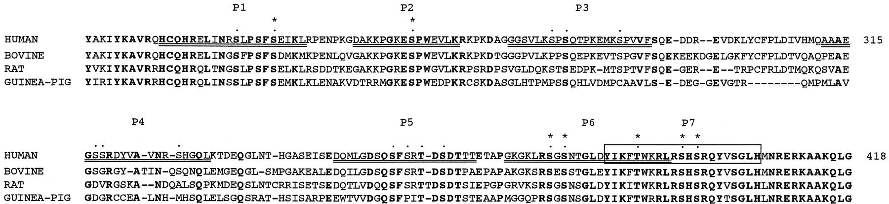
Recent cloning studies of H1Rs from bovine (Yamashita et al., 1991), rat (Fujimoto et al., 1993), guinea pig (Horio et al., 1993; Traiffort et al., 1994), mouse (Inoue et al., 1996), and human (De Backer et al., 1993; Fukui et al., 1994; Moguilevsky et al., 1994) have demonstrated that many threonine and serine residues are present in the third intracellular loop. Figure 1 shows the amino acid sequences of the third intracellular loop of the H1Rs. Many potential phosphorylation sites are located in human H1R that are consistent with the consensus sequence for substrate recognition by PKC (Kennelly and Krebs, 1991). Some are also conserved among other species. To examine whether these amino acids could be phosphorylated by PKC, we synthesized seven peptides (P1 through P7) corresponding to partial sequences in the third intracellular loop of the H1R, and we used these peptides as substrates for in vitro phosphorylation experiments. Of the seven peptides, five were not phosphorylated at all. Of the remaining two peptides (P6 and P7), phosphorylation of P6 by PKC was very weak compared with that of P7 (Fig.2). Of the three potential phosphorylation sites present in P7, Thr390 is also contained in P6, which is only weakly phosphorylated. Thus we speculated that the other two sites (Ser396 and/or Ser398) might be PKC phosphorylation sites.

Putative PKC phosphorylation sites in the third intracellular loop of H1R. Amino acid sequences of the third intracellular loop of human, bovine, rat, and guinea pig H1Rs are shown. Dotted residues indicate putative PKC phosphorylation sites in the third intracellular loop of human H1R, and asterisked residues are putative PKC phosphorylation sites that are conserved among these four species. Synthetic peptides (P1-P7), which are underlined or boxed, were used for in vitro phosphorylation experiments. The amino acid numbers in the sequence are shown to the right of the sequence.

Phosphorylation by PKC of synthetic peptides corresponding to regions of the third intracellular loop of human H1R. In vitro phosphorylation experiments were performed using synthetic peptides (Fig. 1, P1-P7) corresponding to partial sequences of the third intracellular loop of the human H1R. Each peptide (1 μg) was phosphorylated by PKC with 20 μM [γ-32P]ATP for 30 min at 30°C. The reaction products were separated by SDS-PAGE, and then visualized by autoradiography (lanes 1 to 7 corresponding to P1 through P7, respectively).
To determine which residues can be phosphorylated by PKC, we synthesized mutant peptides in which serine residues 396 and 398 were substituted with an alanine residue. As shown in Fig.3A, both peptides P7-S396A (Ser396 to alanine) and P7-S398A (Ser398 to alanine) were phosphorylated by PKC, but the extent of phosphorylation was small compared to the peptide corresponding to the wild-type receptor. P7-S396A/S398A (both Ser396 and Ser398 to alanine) was also phosphorylated by PKC, but the phosphorylation level was lower than that seen with P7-S396A or P7-S398A. Quantitative analysis of wild-type and mutant peptides of the H1R demonstrated that 31% and 45% of the total count was incorporated at Ser396 and Ser398, respectively (Fig. 3B). These data suggest that both serine residues can be phosphorylated by PKC.

Determination of 32P incorporated into Ser396 and Ser398 in human H1R. We compared PKC phosphorylation of Ser396 and Ser398 in human H1R using P-7 peptides of wild-type (lane 1), S396A mutant (lane 2), S398A mutant (lane 3), and S396A/S398A double mutant (lane 4) H1R. Each peptide (1 μg) was phosphorylated by PKC with 20 μM [γ-32P]ATP for 30 min at 30°C. The reaction products were separated by SDS-PAGE and then visualized by autoradiography (A). The signals were quantified by BAS-2000II (B).
Expression of Wild-Type or Mutant H1R in CHO cells.
The pdKCR-dhfr vectors containing wild-type or mutant H1R gene were transfected into dihydrofolate reductase-deficient CHO cells in which H1R was not detectable by [3H]mepyramine binding assay, and transfected cells were selected using medium devoid of nucleosides. Individual colonies were isolated, and expression levels were analyzed by [3H]mepyramine binding assay. Clones expressing the receptor at similar high levels (0.5–1.5 pmol/mg of protein) were selected for further experiments. The pharmacological properties of wild-type or mutant H1R expressed in the selected clones were shown in Table 1. The affinities for [3H]mepyramine and histamine of the wild-type H1R were consistent with the reported values (Haaksma et al., 1990; Hill, 1990). The Kd values for [3H]mepyramine and theKi values for histamine were not significantly altered in any of the mutant H1Rs.
Binding properties of wild-type and mutant H1R expressed in CHO cells. Plasma membranes from CHO cells expressing wild-type or mutant H1R were prepared, and [3H]mepyramine binding assays were performed as described in Experimental Procedures. Kdand Bmax values for [3H]mepyramine binding were obtained from Scatchard plots. Results represent the mean ± standard error of three independent experiments.
Desensitization of Wild-Type or Mutant H1R by PMA.
We examined the effect of PMA on histamine-induced accumulation of IP in CHO cells expressing wild-type H1R. Histamine-induced IP accumulation could be observed for 60 min after histamine treatment, and this accumulation was reduced in the presence of PMA (Fig.4A). PMA attenuated histamine-induced accumulation of IP (Fig. 4B) in a dose-dependent manner. Treatment with 100 nM PMA induced maximal desensitization, which was 26% of the accumulation of IP seen in the control. The EC50value was 6.2 nM, which was comparable with that for PMA-induced PKC activation (Castagna et al., 1982). Pretreatment of these cells with K252a and staurosporine, inhibitors of PKC, led to a dose-dependent increase of the histamine-induced accumulation of IP from the PMA-induced desensitized state to the nondesensitized state (Fig. 4C). Pretreatment with 1 μM staurosporine could completely block the inhibitory effect of PMA, and the IC50 value of 89 nM was comparable with that for the inhibition of PKC activity. The IC50 ratio for staurosporine versus K252a is 1/10, which agrees with the calculated ratio from the reportedKi values of the two inhibitors for PKC. These results suggested that PMA-induced PKC activation caused desensitization of H1R-mediated accumulation of IP in CHO cells.

Effect of PKC activation on histamine-induced IP accumulation in CHO cells expressing wild-type H1R. A, time course of histamine-induced accumulation of IP. CHO cells expressing wild-type H1R were labeled with myo-[3H]inositol for 24 h and then treated with 100 μM histamine in the presence of Li+ for different periods of time in the absence (E) or presence (J) of 1 μM PMA. The results are presented as increase in IP accumulation above the basal levels in the absence of histamine. B, dose-response to PMA of histamine-induced accumulation of IP. [3H]Inositol-labeled cells were treated with 100 μM histamine in the presence of Li+ for 20 min with the indicated concentrations of PMA. The histamine-induced accumulation of IP in the absence of PMA represents 100%. C, effect of PKC inhibitors on PMA-induced desensitization of human H1R. [3H]Inositol-labeled cells were pretreated with the indicated concentrations of K252a (E) or staurosporine (J), and were stimulated with 100 μM histamine in the presence of Li+and 1 μM PMA for 20 min. The histamine-induced accumulation of IP in the absence or presence of PMA represents 100% and 0%, respectively. The data represent the mean ± standard error of triplicate values from three separate experiments.
To determine whether Ser396 and/or Ser398 are involved in the desensitization of H1R by PMA, we examined the effects of PMA on histamine-induced accumulation of IP in CHO cells expressing mutant H1R or wild-type H1R. Treatment with 10 μM histamine induced IP formation in a time dependent manner in CHO cells expressing wild-type H1R, and this IP formation was almost completely abolished by 1 μM PMA pretreatment (Fig. 5A). In cells expressing the S396A mutant H1R, IP formation was reduced to 70% of the control level in the presence of PMA within 5 min of 10 μM histamine treatment, but further accumulation of IP was not observed after longer exposure of the cells to histamine with PMA (Fig. 5B). In cells expressing the S398A or S396A/S398A mutant H1Rs, time-dependent IP formation was induced by histamine in the presence of PMA, and IP formation remained at 80 to 85% of the control level (Fig. 5, C and D).

Time course of histamine-induced accumulation of IP in CHO cells expressing wild-type or mutant H1R. CHO cells expressing wild-type or mutant H1R were labeled with myo-[3H]inositol for 24 h, and then treated with 10 μM histamine in the presence of Li+ for different periods of time in the absence (E) or presence (J) of 1 μM PMA. The data represent the mean ± standard error of triplicate values from three separate experiments.
We investigated the ability of histamine with or without PMA to activate PLC mediated by wild-type or mutant H1R (Fig.6). In cells expressing wild-type H1R, accumulation of IP increased in a dose-dependent manner after histamine treatment, and this histamine-induced accumulation of IP was desensitized by 1 μM PMA treatment (Fig. 6A). Weak desensitization (maximal response of IP reduced by 15–20% compared with controls) was observed in S398A and S396A/S398A mutants (Fig. 6, C and D, and Table2), whereas strong desensitization (maximal IP levels reduced by 50–65%) was observed in wild-type and the S396A mutant, respectively (Fig. 6, A and B, Table 2). Thus, substitution of alanine for Ser398 considerably decreased PMA-induced desensitization of the H1R. On the other hand, substitution of alanine for Ser396 had less effect; desensitization of the double mutant was not significantly different from that of the S398A mutant H1R.

Histamine dose-response for histamine-induced accumulation of IP in CHO cells expressing wild-type or mutant H1R. CHO cells expressing wild-type or mutant H1R were labeled withmyo-[3H]inositol for 24 h, and then treated with the indicated concentrations of histamine in the presence of Li+ for 20 min in the absence (E) or presence (J) of 1 μM PMA. The data represent the mean ± standard error of triplicate values from three separate experiments.
Comparison of EC50 values and maximal responses for histamine-induced inositol phosphates accumulation in CHO cells expressing wild-type or mutant H1R.
We examined EC50 values for histamine-induced IP accumulation in CHO cells expressing wild-type or mutant H1R (Table 2). Treatment with PMA induced a 10-fold increase in the EC50 value in the wild-type H1R. In the S396A mutant, a marked increase (6-fold) in the EC50value was observed after treatment with PMA, although the extent was slightly smaller than that in the wild-type. On the other hand, changes in the EC50 values were very small for the S398A and S396A/S398A mutants (2-fold and 1.5-fold, respectively). Both the S398A and S396A/S398A mutants lack the putative PKC phosphorylation site (Ser398). In the absence of PMA, the EC50 values of the S398A and S396A/S398A mutant H1Rs were 3.5- to 4.5-fold lower than those of the wild-type and S396A mutant H1Rs. To determine whether the difference in the EC50 values results from endogenous phosphorylation of Ser398 or from structural changes brought about by substitution of alanine for Ser398, we examined the effect of 1 μM staurosporine, which could largely block endogenous PKC activity, on histamine-induced accumulation of IP. Treatment with staurosporine resulted in a 2-fold decrease in the EC50 value with no change inVmax for histamine-induced accumulation of IP in CHO cells expressing wild-type H1R but did not significantly change these values in the S396A/S398A mutant. As a result, the difference in the EC50 values between wild-type and S396A/S398A mutant H1R was reduced from 3.9-fold to 1.4-fold. These results strongly suggest that Ser398 is partially phosphorylated by endogenous PKC in the absence of PMA and that this phosphorylation is responsible for the increase in the EC50 value for histamine-induced accumulation of IP in CHO cells expressing wild-type H1R.
Discussion
Phorbol ester-induced desensitization of the H1R seems to be dependent on PKC activation. However, little is known about the target proteins phosphorylated by PKC. Recent studies on receptor desensitization have demonstrated that receptor phosphorylation is a key event in the desensitization mechanism. Post and Dawson have reported that PKC activation inhibits histamine-induced accumulation of IP but does not affect IP accumulation induced by the nonspecific G protein activator AF in oligodendroglioma cells (Post and Dawson, 1992), which indicates that the target protein is probably at the receptor level. In the present study, our data strongly suggests that direct phosphorylation of the H1R is involved in PKC-mediated desensitization. Two amino acids (Ser396, Ser398) were phosphorylated by PKC in vitro. Although there is no direct evidence to suggest that phorbol ester-induced PKC activation induces phosphorylation of these amino acids in the native protein, site-directed mutagenesis studies indicated that these serine residues, particularly Ser398, are involved in phorbol ester-induced desensitization. Production of antibody against H1R protein may be useful to demonstrate the direct phosphorylation of H1R by PKC.
PKC consists of a family of isoenzymes. These isoenzymes show differential tissue expression and specific intracellular localization. PKC phosphorylates its target proteins at specific sites. However, there have been no reports on differences in substrate specificity between the isoenzymes. Furthermore, it is not known which PKC isoenzymes are physiologically involved in phosphorylation of the H1R. Therefore, a mixture of α, β, and γ-PKC was used in the present experiment for the phosphorylation of synthetic peptides corresponding to partial sequences of the third intracellular loop of the H1R. A large number of studies have shown that PKC recognizes the motif (Arg/Lys)1–3-X0–2-Ser/Thr-X0–2-(Arg/Lys)1–3> Ser/Thr-X0–2-(Arg/Lys)1–3≥ (Arg/Lys)1–3-X0–2-Ser/Thr (where X can be any amino acid residue) (Kennelly and Krebs, 1991). We previously reported that two potential PKC phosphorylation sites (Ser395, Ser397) are present in the third intracellular loop of rat H1R (Fujimoto et al., 1993). These sites were conserved in bovine (Yamashita et al., 1991), guinea pig (Horio et al., 1993; Traiffort et al., 1994), mouse (Inoue et al., 1996) and human H1R (De Backer et al., 1993; Fukui et al., 1994; Moguilevsky et al., 1994). In the present study, we have shown that these two amino acid residues (human H1R; Ser396, Ser398) could be phosphorylated by PKC. On the other hand, potential phosphorylation sites for PKC in the third intracellular loop of the H1R that have a sequence that fits the consensus motif less well were not phosphorylated by PKC. Ser396 and Ser398 are also potential phosphorylation sites for cAPK, cGMP-dependent protein kinase, and CaMK II. Elevation of intracellular cAMP content has been shown to attenuate histamine-induced accumulation of IP in C6 glioma cells (Peakman and Hill, 1994) and in DDT1 MF-2 smooth muscle cells (Sipma et al., 1995). cAMP-induced H1R desensitization may also result from phosphorylation of these serine residues by cAPK. In addition, the possibility exists that cGMP-dependent protein kinase and CaMK II are also involved in desensitization of the H1R.
Treatment with PMA resulted in attenuation of histamine-induced accumulation of IP in CHO cells expressing wild-type H1R. PMA-induced desensitization was abolished by pretreatment with K252a or staurosporine, PKC inhibitors, or by down-regulation of PKC by prolonged treatment with PMA (data not shown). These results demonstrate that PKC activation is required for desensitization of the H1R, in agreement with previous reports (McCreath et al., 1994; Zamani et al., 1995). Our data demonstrated that the inhibitory effect of PMA was partial (maximal decrease was about 70% with 100 nM PMA), whereas previous studies have shown that PMA almost completely inhibits histamine-mediated signaling in various cell lines that endogenously express the H1R (Kotlikoff et al., 1987; Smit et al., 1992; Daykin et al., 1993; Zamani et al., 1995). This deficiency in the effect of PMA may be a result of the high level of expression of the H1R, because similar results have also been observed in cells stably transfected with the H1R gene (Mizuguchi et al., 1994; Smit et al., 1996). Treatment with PMA induced a 10-fold increase in the EC50 value for histamine-induced accumulation of IP in CHO cells expressing wild-type receptors, which implies that phosphorylation of the H1Rs by PKC caused G-protein uncoupling. This agrees with previous reports that receptor-G protein uncoupling is involved in the desensitization process (Yuan et al., 1994; Ng et al., 1995). Pretreatment with staurosporine caused a 2-fold decrease in the EC50 value, which suggests that the H1R is partially phosphorylated by basal PKC activity in the absence of PMA and this phosphorylation is related to the decline in sensitivity to histamine.
We constructed mutant receptors, in which Ser396 and/or Ser398 were replaced by alanine, using the techniques of site-directed mutagenesis. CHO cells expressing wild-type and mutant receptors at similar levels were selected by [3H]mepyramine binding and were characterized. In the absence of PMA, the ability of histamine to activate PLC was similar in both wild-type and mutant H1Rs, whereas the effect of pretreatment with PMA on histamine-induced accumulation of IP was different between the wild-type and mutant H1Rs. PMA-induced desensitization was significantly suppressed by substitution of alanine for Ser398, but was reduced less by substitution of alanine for Ser396, which indicates that Ser398 is the more important site for PMA-induced desensitization. These serine residues are located in the carboxyl-terminal domain of the third intracellular loop. It has been shown that this domain is important for coupling to the G-protein in the muscarinic receptor (Burstein et al., 1995).
These mutations of the H1R did not affect ligand binding (Kd for [3H]mepyramine binding andKi for histamine binding). The S398A mutation caused a leftward shift in the EC50value for histamine-induced accumulation of IP compared with the wild-type receptor. This change cannot be attributed to a conformational change; rather, it is caused by a difference in the endogenous level of phosphorylation between the two receptors, because treatment with staurosporine significantly decreases the difference in the EC50 values between the wild-type and S396A/S398A mutant.
In the S398A and S396/398A mutant receptors, PMA-induced desensitization was largely abolished (as in the wild-type receptor), but slight desensitization remained. Another mechanism (for example, PKC phosphorylation of downstream molecules or phosphorylation of H1R at sites other than Ser396 and Ser398) may also be involved in PMA-induced desensitization. Further potential PKC phosphorylation sites are present in the first intracellular loop and carboxyl terminus of H1R (Horio et al., 1993). Murray et al. (1989)have reported that PMA causes a reduction in PLC activity, possibly mediating in part a desensitization of histamine-induced IP3 formation. In addition, Tilly et al. have reported that PKC activation attenuates guanosine-5′-O-(3-thio) triphosphate-induced IP formation (Tilly et al., 1990). The mutants described here will be useful for further studies to determine whether any of these mechanisms are involved in PMA-induced desensitization.
In the present study, we have shown that treatment with PMA induces uncoupling of the H1R from associated G proteins, leading to attenuation of histamine induced-accumulation of IP in CHO cells expressing the H1R. In vitro phosphorylation experiments have demonstrated that two amino acid residues (Ser396, Ser398) in the third intracellular loop of the H1R could be phosphorylated by PKC. Site-directed mutagenesis studies suggest that phosphorylation of Ser398 plays an important role in the PMA-induced desensitization of the H1R.
Acknowledgments
We thank Drs. S. Ito, R. Yoshida and O. Hayaishi for expert advice with this project. We also thank Drs. H. Shimono and M. Yabumoto for encouragement.
Footnotes
-
Send reprint requests to: Dr. Hiroyuki Fukui, Department of Pharmacology, Faculty of Pharmaceutical Sciences, University of Tokushima, 1–78-1 Shomachi, Tokushima 770-8505, Japan. E-mail:hfukui{at}ph.tokushima-u.ac.jp
-
↵1 Current affiliation: Dept. of Biochemistry, School of Dentistry, Hiroshima University, Hiroshima, Japan.
-
This work was supported in part by grants from JSPS Research Fellowships and Research Foundation for Clinical Pharmacology, and Grants-in-Aid from the Ministry of Education Science and Culture of Japan.
- Abbreviations:
- H1R
- histamine H1 receptor
- PLC
- phospholipase C
- IP3
- inositol 1,4,5-trisphosphate
- PMA
- phorbol-12-myristate-13-acetate, PKC, protein kinase C
- cAPK
- cAMP-dependent protein kinase
- CaMK II
- calcium/calmodulin-dependent protein kinase II
- GRK
- G protein-coupled receptor kinase
- CHO
- Chinese hamster ovary
- IP
- inositol phospate(s)
- SDS
- sodium dodecyl sulfate
- PAGE
- polyacrylamide gel electrophoresis
- Received October 1, 1998.
- Accepted December 17, 1998.
- The American Society for Pharmacology and Experimental Therapeutics


