


Abstract
P38α is a protein kinase that regulates the expression of inflammatory cytokines, suggesting a role in the pathogenesis of diseases such as rheumatoid arthritis (RA) or systemic lupus erythematosus. Here, we describe the preclinical pharmacology of pamapimod, a novel p38 mitogen-activated protein kinase inhibitor. Pamapimod inhibited p38α and p38β enzymatic activity, with IC50 values of 0.014 ± 0.002 and 0.48 ± 0.04 μM, respectively. There was no activity against p38δ or p38γ isoforms. When profiled across 350 kinases, pamapimod bound only to four kinases in addition to p38. Cellular potency was assessed using phosphorylation of heat shock protein-27 and c-Jun as selective readouts for p38 and c-Jun NH2-terminal kinase (JNK), respectively. Pamapimod inhibited p38 (IC50, 0.06 μM), but inhibition of JNK was not detected. Pamapimod also inhibited lipopolysaccharide (LPS)-stimulated tumor necrosis factor (TNF) α production by monocytes, interleukin (IL)-1β production in human whole blood, and spontaneous TNFα production by synovial explants from RA patients. LPS- and TNFα-stimulated production of TNFα and IL-6 in rodents also was inhibited by pamapimod. In murine collagen-induced arthritis, pamapimod reduced clinical signs of inflammation and bone loss at 50 mg/kg or greater. In a rat model of hyperalgesia, pamapimod increased tolerance to pressure in a dose-dependent manner, suggesting an important role of p38 in pain associated with inflammation. Finally, an analog of pamapimod that has equivalent potency and selectivity inhibited renal disease in lupus-prone MRL/lpr mice. Our study demonstrates that pamapimod is a potent, selective inhibitor of p38α with the ability to inhibit the signs and symptoms of RA and other autoimmune diseases.
Diseases of chronic inflammation, such as rheumatoid arthritis (RA), psoriatic arthritis, and inflammatory bowel disease, are driven by the release of proinflammatory cytokines and other mediators that disrupt normal tissue integrity. The synthesis and release of these mediators are controlled by various signaling pathways that are integrated largely by the mitogen-activated protein kinases (MAPKs). One of these MAPKs, p38, has been implicated in the regulation of a variety of immune cell functions (Schieven, 2005). A range of stimuli can activate p38, including toll-like receptor ligands, TNF-α, and IL-1 and cellular stresses, such as heat shock and hypoxia (Chen et al., 2001; Karin, 2005). Upon activation, p38 regulates a range of genes that are implicated in the inflammatory response, such as TNF-α, IL-1, IL-6, and many others (Zhang et al., 2007).
The role of p38 in response to and regulation of inflammatory pathways has led to efforts to develop inhibitors of p38 as therapeutic agents for diseases characterized by chronic inflammation (Kumar et al., 2003; Goldstein and Gabriel, 2005; Westra and Limburg, 2006). Involvement of p38 in the pathogenesis of RA is indicated by its activation in the synovial tissue of RA patients (Schett et al., 1998; Inoue et al., 2006). The potential of p38 inhibitors to inhibit human disease progression is suggested by their efficacy in rodent models of arthritis (Badger et al., 1996; Nishikawa et al., 2003; Wada et al., 2005; Medicherla et al., 2006). In addition, based on the role of p38 in secretion of TNF and the success of anti-TNF therapies in RA, several p38 inhibitors have been tested in clinical trials as a therapy for RA. It is unfortunate that poor safety profiles have been observed in phase II clinical trials because of adverse effects on the central nervous system and liver (Zhang et al., 2007). To date, the presence of these dose-limiting toxicities has prevented adequate exploration of efficacy, which underscores the importance of separating p38-driven effects from the potentially toxic off-target effects of these compounds.
The specific effects of p38 inhibition have been investigated using both murine knockout models and pharmacologic inhibitors of p38. There are four isoforms of p38 (α, β, δ, γ) that demonstrate differential tissue expression (Hale et al., 1999; Alonso et al., 2000; Court et al., 2002) and activation (Alonso et al., 2000). The commonly used (but not selective) p38 inhibitors, SB203580 and SB202190, have equivalent potency against both the α and β isoforms but do not inhibit the δ and γ isoforms (Kumar et al., 2003; Saklatvala, 2004). These compounds are effective inhibitors of inflammation, indicating the importance of p38α and/or β in inflammatory responses. Mice deficient in p38β, however, are viable and healthy and showed normal downstream signaling, immediate early gene transcription, lipopolysaccharide (LPS)-induced cytokine production, and T cell development. In addition, these mice remained susceptible to inflammatory disease when crossed onto the TNFα-overexpressing TNFΔARE mouse line (Beardmore et al., 2005). In contrast, mice deficient in p38α are embryonic lethal because of a placental defect (Adams et al., 2000; Allen et al., 2000; Mudgett et al., 2000). Taken together, these data are consistent with the α isoform being the major contributor to the role of p38 in inflammation.
In addition to the effects of p38 inhibitors on various p38 isoforms, selectivity against other protein kinases is an important consideration in understanding the biology of p38 inhibition. Because most kinase inhibitors in development target the binding site for the common substrate ATP, designing a selective ligand requires the identification of unique interaction sites around the ATP binding pocket. Discerning the degree of selectivity depends on the number of different kinases that can be screened with a compound. In addition, despite the availability of technologies that allow screening of large numbers of kinases (Fabian et al., 2005), these technologies do not allow a comparable level of selectivity assessment in cells in which cellular context can change the efficacy of inhibitors dramatically. This limitation has important implications for our understanding of p38 biology because the compounds that have been used widely to test the effects of p38 inhibition on various cell types are not completely selective in vitro (Fabian et al., 2005); furthermore, their selectivity in cells has not been characterized.
In the current study, we present evidence that the p38 inhibitor pamapimod is highly selective both in vitro and in cell culture. In addition, the preclinical data support the conclusion that pamapimod has the ability to inhibit the signs and symptoms of RA and other autoimmune disease.
Materials and Methods
Kinase Isoform Selectivity Assays. Purified recombinant human active p38α, β, γ, or δ (10 nM) diluted in 20 mM MOPS, pH 7.2, 25 mM β-glycerophosphate, 5 mM EGTA, 1 mM dithiothreitol, 1 mM sodium orthovanadate, and 40 mM MgCl2 was mixed with 10× pamapimod at a final concentration of 100 to 0.0003 μM in a 96-well plate (Greiner Bio-One, Longwood, FL) and incubated for 10 min at room temperature. The kinase reaction was initiated by the addition of 10 μl of 4× substrate cocktail containing Km concentrations of the substrate recombinant myelin basic protein (30 μM; Invitrogen, Carlsbad, CA), ATP (40 μM; Roche Applied Science, Indianapolis, IN), and γ-[33P]ATP (2 μCi/rxn; Amersham Inc., Chalfont St. Giles, UK). After a 30-min incubation at 30°C, the kinase reaction was terminated by the transfer of 25 μl of the reaction mixture to a phosphocellulose membrane/plate (Millipore Corporation, Billerica, MA) containing 150 μl of 0.75% phosphoric acid (Mallinckrodt Baker, Inc., Phillipsburg, NJ). The next day, the free nucleotides in the membrane were washed under vacuum with 3 × 200 μl of 0.75% phosphoric acid. After the last wash, membrane/plates were transferred to an adaptor plate (PerkinElmer Life and Analytical Sciences, Waltham, MA), and 70 μl of scintillation cocktail (PerkinElmer Life and Analytical Sciences) was added to each well. After at least 4 h, signal development the amount of radioactivity was counted on a top counter.
Inhibition of TNF-α Secretion by a Human Monocytic Cell Line, THP-1. THP-1 cells, growing in log phase, were collected by centrifugation and resuspended in RPMI 1640 (Invitrogen) containing 5.5 × 10⁻⁵ M 2-mercaptoethanol (Invitrogen), and 10% fetal bovine serum (Summit Biotechnology, Fort Collins, CO) to a final cell concentration of 2.5 × 10⁶ cells/ml. Dilutions of pamapimod were predispensed in 25-μl aliquots (before addition of cells) into round-bottom 96-well plates (U-bottom TC plate, no. 3799; Corning Life Sciences, Lowell, MA). The starting concentration was 100 μM in 5% dimethyl sulfoxide, and six half-log serial dilutions were made. After the addition of 200 μl of cell suspension and 25 μl of 5 μg/ml LPS (Escherichia coli, 0127, B8; Sigma-Aldrich, St. Louis, MO) in medium, the final dimethyl sulfoxide concentration was 0.5%. Compounds were diluted an additional 10-fold, and the final LPS concentration was 500 ng/ml. The cell suspensions and compound dilutions were combined and incubated for 30 min at 37°C in a 5% CO2 humidified atmosphere, before the addition of LPS (or medium for non-LPS control samples). After the addition of LPS, plates were incubated for 2 h, followed by centrifugation to pellet cells. Cell supernatants were stored at 4°C until analysis for TNF-α content. TNF-α levels were determined by ELISA (Antibody Solutions, Mountain View, CA) following the manufacturer's directions. Cytokine concentrations were determined from a standard curve using Soft-Max Pro (Molecular Devices, Sunnyvale, CA). The percentage inhibition was calculated for each pamapimod concentration tested, and an IC50 curve was constructed using Xlfit software.
Inhibition of IL-1β Biosynthesis in LPS-Stimulated Human Whole Blood. Blood was collected from healthy volunteers (drug-free for 2 weeks) into Vacutainers (BD Biosciences, San Jose, CA) containing 19 units/ml sodium heparin. Pamapimod was predispensed and diluted as described for the THP-1 assay above. Human whole blood (200 μl/sample) was preincubated with the diluted compound for 30 min at 37°C, followed by the addition of 25 μl of LPS dissolved in RPMI 1640 (Invitrogen) to produce a final LPS concentration of 0.5 μg/ml. Negative control wells received RPMI 1640 alone. Plates were incubated for 18 h at 37°C in a 5% CO2 atmosphere followed by centrifugation at 150g for 10 min. Plasma was collected from each sample and stored in polypropylene plates at 4°C. For IL-1β determinations, plasma was diluted 4-fold in ELISA diluent buffer and tested by ELISA (Antibody Solutions) following the manufacturer's instructions. Cytokine levels were detected and calculated as described above for the THP-1 assay.
Detection of p38 and JNK Activity in Cells or Tissue Samples by Western Blotting. Selectivity of inhibition of p38 and JNK in a cellular context was determined by measuring phosphorylation of HSP-27 and c-Jun, respectively, in SW-1353 or THP-1 cells. After 30-min pretreatment with pamapimod, cells were stimulated with TNFα (R&D Systems, Minneapolis, MN) for 20 min and then rinsed with ice-cold phosphate-buffered saline and lysed in 100 μl of ice-cold lysis buffer (20 mM Tris, pH 7.5, 150 mM NaCl, 1 mM EDTA, 1 mM EGTA, 1% Triton X-100, 2.5 mM sodium pyrophosphate, 1 mM β-glycerophosphate, and 1 mM Na3V04; Cell Signaling Technology Inc., Danvers, MA) plus protease inhibitor mixture (Roche Applied Science). After sonicating 10 times for 1 s each on ice, the samples were centrifuged at 12,000g for 5 min at 4°C. The protein concentration was measured, and 15 μg of protein from each sample was separated by SDS-polyacrylamide gel electrophoresis and transferred to nitrocellulose membranes. Membranes were blocked in TBST buffer (20 mM Tris, pH 7.5, 136 mM NaCl, 0.1% Tween 20) containing 5% milk and incubated with the primary antibodies for phospho-HSP27, phospho-c-Jun, phospho-p38, p38, HSP27 (1:1000 dilution in TBST containing 5% bovine serum albumin; Cell Signaling Technology Inc.), and c-Jun (1:1000 dilution in TBST containing 5% bovine serum albumin; Millipore Bioscience Reagents, Temecula, CA). Western blots of cell lysates were analyzed by the addition of IR Dye 700 Dx goat anti-rabbit and IR Dye 800 goat anti-mouse antibodies (Rockland Immunochemicals, Gilbertsville, PA), and the multicolor fluorescence was detected with the Odyssey imaging system (LI-COR Biosciences, Inc., Lincoln, NE). Density of phosphoprotein bands was measured using ImageQuant software (Amersham Inc.). The percentage inhibition of pamapimod-treated phosphoprotein, normalized with level of total protein, was calculated and an IC50 curve was constructed using Xlfit software.
To demonstrate modulation of p38 activity in vivo, phosphorylation of HSP-25 in tissues from either DBA/1J (murine collagen-induced arthritis model) or MRL/lpr (lupus model) mice was assessed by Western blotting after dosing with pamapimod. Tissue lysates from lung or inguinal lymph nodes were prepared by homogenization in 10 mM Tris, 100 mM NaCl, 1 mM EDTA, 1 mM EGTA, 1 mM sodium fluoride, 20 mM sodium phosphate, 2 mM sodium vanadate, 1% Triton X-100, 10% glycerol, 0.1% SDS, and 0.5% deoxycholate (BioSource International, Camarillo, CA) with protease inhibitors. Western blots were probed for total HSP-25 (Santa Cruz Biotechnology, Inc., Santa Cruz, CA), phospho-HSP-25 (BioSource International), total p38, and phospho-p38 (Cell Signaling Technology Inc.) and analyzed either by the addition of horseradish peroxidase-coupled secondary antibodies and developed with the enhanced chemiluminescence detection system (Amersham Inc.) or by the addition of Alexa Fluor 647 goat anti-mouse (Invitrogen), and the multicolor fluorescence was detected with the Typhoon imaging system (Amersham Inc.).
Murine Acute LPS-Induced Cytokine Production Models. All animal procedures were approved by and conducted in accordance with the Roche Palo Alto Institutional Animal Care and Use Committee guidelines. Female BALB/c mice (The Jackson Laboratory, Bar Harbor, ME) were utilized for in vivo LPS or TNF-α stimulation studies. In all experiments, each dose group consisted of eight animals (each 18–25 g in weight). The test materials were administered in a volume of 1 ml/100 g body weight as suspensions in an aqueous vehicle containing 0.9% NaCl, 0.5% sodium carboxymethylcellulose, 0.4% polysorbate 80, 0.9% benzyl alcohol, and 97.3% distilled water. Mice were orally administered vehicle or the indicated doses of pamapimod 30 min before LPS (E. coli, serotype 0127:b8; Sigma-Aldrich) or TNF-α intraperitoneal challenge. LPS was administered to mice at 800 μg/kg. Animals were euthanized via CO2 inhalation 1.5 h after LPS or TNF-α administration, and blood was collected and transferred to SST Vacutainer serum separator tubes. The sera were separated and stored at -20°C until analysis by ELISA. ELISA kits for TNF-α and IL-6 were obtained from BioSource International. All ELISAs were performed according to the manufacturer's specifications.
Brewer's Yeast-Induced Paw Hyperalgesia Model. To measure effects of p38 inhibition on inflammatory pain, we utilized the Randall-Selitto test of mechanical hyperalgesia. Male Sprague-Dawley rats (115–150 g; Charles River Laboratories, Inc., Wilmington, MA) were acclimated for 1 week and randomized into groups of 10 animals each. A solution of 20% Brewer's yeast (0.1 ml; Sigma-Aldrich) was injected into the left hindpaw of each animal at the beginning of the study. Two hours later, rats were orally administered vehicle (1 ml/kg), indomethacin (5.0 mg/kg), or pamapimod at doses of 10 to 100 mg/kg. After an additional 1 h, the left hindpaw of each rat was placed on the platform of the Basile Analgesy-meter, and the mechanical force in grams at which the rat withdraws its paw was recorded. After the assay, the animals were euthanized via CO2 asphyxiation and bled for serum collection.
Murine Collagen-Induced Arthritis Model. DBA/1J female mice (The Jackson Laboratory) were between 8 and 10 weeks of age at the initiation of the experiments. Each dose group consisted of 12 animals. Test materials were administered in a volume of 0.2 ml in the aqueous vehicle described above. On day 0, mice were injected intradermally at the base of the tail with 0.1 ml of an emulsion of Bovine Type II Collagen (100 μg; Chondrex, Inc., Redmond, WA) and an equal volume of Complete Freund's adjuvant (H37 RA; Difco, Detroit, MI). Approximately 4 weeks after collagen injection, the animals were challenged with an intraperitoneal injection of LPS (50 μg). Mice were scored daily after LPS challenge for the development of arthritis in paws until disease became evident (mean clinical score = 3–4). After detection of disease in the majority of animals, the mice were randomized into dosing groups, and treatment was initiated. Mice displaying no arthritis or disease outside worse than the target clinical score were removed from the studies. Pamapimod and vehicle were administered by oral gavage (0.2 ml) once daily. Scoring was as follows: 1, swelling and/or redness of paw or one digit; 2, swelling in two or more joints; 3, gross swelling of the paw with more than two joints involved; and 4, severe arthritis of the entire paw and digits. The arthritic index for each mouse is determined by adding the four scores of the individual paws.
To measure effects on bone destruction, ex vivo micro-CT imaging and analysis were performed on the calcaneus, tarsal bones, and phalanges of formaldehyde-fixed paw samples. Regions of interest were hand-drawn over the reconstructed CT images of the calcaneus and tarsal bones. A 2.1-mm-diameter region of interest was centered over the metatarsal-phalangeal joint of each toe. The bone volume was calculated from each of these regions of the paw. The bone volumes did not include the lower density periosteal bone formed in diseased tissue.
Histopathology of Kidneys from MRL/MpJ-Faslpr/J (MRL/lpr) Mice. MRL/MpJ-Faslpr/J (MRL/lpr) mice from The Jackson Laboratory were maintained under specific pathogen-free conditions. Necropsy was performed on sacrificed mice, and one kidney from each animal was fixed in 10% neutral-buffered formalin. The kidney was cross-sectioned into four quadrants, with the first sections made on either side of the renal papilla and the second sections made lateral to the first. This method enabled review of each renal pole and the renal papilla with arcuate vessels. Sections were paraffin-embedded, sectioned at 5 μm thick, stained with hematoxylin and eosin, and reviewed by light microscopy. In a subset of animals, serial 3-μm-thick sections were stained with periodic acid-Schiff stain for mucopolysaccharide-based material (including fibrin and glomerular deposition of noncollagenous material). The histopathology parameters evaluated were arteritis, glomerulonephritis, and perivascular mononuclear infiltration. Arteritis was recorded as the total number of vessels affected across the four sections to the exclusion of the juxtaglomerular apparatus. Glomerulonephritis was recorded as 1 to 5 according to the most severe morphologic pattern identified from one of the five morphologic patterns of lupus glomerulonephritis as defined by the World Health Organization. The perivascular mononuclear infiltrate was scored as 1 to 5 according to the average from all vessels across all four sections and based on the average width of the band between the vein and proximate arteriole or in the papilla, as the average width of the band between the renal vein and urinary space and interlobular vein and artery (1, minimal or few leukocytes; 2, mild or <1× tubular diameter; 3, moderate or ≤2× tubular diameter; 4, marked or >2×-4× tubular diameter; 5, severe or >4× tubular diameter).
Cultured Synovial Explants from Rheumatoid Arthritis Patients. Synovium from patients with rheumatoid arthritis undergoing joint surgery was dissociated by cutting into small pieces, followed by digestion with collagenase type IV and DNase I (Sigma-Aldrich). The mixed cell suspension was cultured for 2 days in the presence or absence of pamapimod at 1 to 2 × 10⁶ cells/ml in RPMI 1640 containing 5% heat-inactivated fetal calf serum, 2 mM l-glutamine, and antibiotics. Supernatants were removed and assayed by ELISA for TNFα.
Results
Kinase Selectivity Profiling of Pamapimod. We initially sought to characterize the selectivity of pamapimod, which is a typical ATP competitive kinase inhibitor that binds the hinge region and interacts with both the front and back pockets of the ATP binding site of p38. The selectivity of pamapimod against a panel of 350 kinases was assessed using the ATP binding site competition assay developed by Ambit Biosciences (San Diego, CA). This technology utilizes phage displayed kinases that are immobilized with nonselective inhibitors. Elution of these immobilized kinases with test compounds reflects the affinity of the compound for a given binding domain. As demonstrated in Fig. 1, pamapimod was quite selective and, other than p38, interacted mainly with JNK isoforms, which similar to p38 are members of the cyclin-dependent kinase, MAPK, glycogen synthetase kinase 3, Cdk2-like kinase family (Table 1) (Manning et al., 2002). In addition, pamapimod interacted with one member of the casein kinase family, Nemo-like kinase, and two isoforms of the ribosomal S6 kinase. Table 2 shows the activity of pamapimod against the four p38 isoforms. Similar to previous p38 inhibitors that have been developed, pamapimod preferentially inhibits the α and β isoforms without activity against the δ or γ isoforms. Table 1 presents Kd values for the kinases from the Ambit screen that showed greater than 85% inhibition at 10 μM concentration. These data indicate that inhibition of the Jnk isoforms could occur in the same concentration ranges as p38 inhibition.
Selectivity of pamapimod determined by Ambit profiling
p38 Isoform selectivity of pamapimod Preactivated recombinant p38 isoforms were assayed for activity by 33P incorporation into myelin basic protein substrate in the presence of varying concentrations of pamapimod as described.

Kinase profiling of pamapimod. A, pamapimod was profiled against 350 kinases at Ambit Biosciences as described under Materials and Methods. Circle size is proportional to percentage inhibition at the test concentration (10 μM). Largest circle, 99% inhibition; medium circle, 90 to 99% inhibition; smallest circles, 50 to 90% inhibition, discounted because this typically translates to a Kd of greater than 1 μM. The kinase dendrogram was adapted from Manning et al. (2002) and is reproduced with permission from Science (http://www.sciencemag.org) and Cell Signaling Technology Inc. (http://www.cellsignal.com). Individual kinase names can be visualized by magnification in PDF format. B, structure of pamapimod.
Because both p38 and JNK are implicated in inflammatory responses, it was necessary to determine whether the activity of pamapimod against p38 and JNK activities detected by phage display would be reflected in cellular activity as well. To assess this, multiplexed substrate phosphorylation assays were performed. Human bone chondrosarcoma cells (SW-1353) were stimulated with TNF, and phospho-c-Jun and phospho-Hsp27 were quantified by Western blots as a reflection of cellular JNK and p38 activity, respectively (Fig. 2). Despite the relatively high affinity of JNK for pamapimod in Ambit profiling, pamapimod was not potent (IC50 > 10 μM) in cells as an inhibitor of JNK. Similar results were obtained using TNF-α stimulation of the monocytic cell line THP-1 (Supplemental Fig. 1). These findings underscore the importance of using cell-based assays to confirm selectivity demonstrated by cell-free binding methods. In addition, it established that the anti-inflammatory activities of pamapimod are likely to occur through a p38 mechanism.
Anti-Inflammatory Activities of Pamapimod. Next, the effects of pamapimod on cytokine production were studied in vitro and in vivo. We focused on the production of TNFα, IL-1β, and IL-6 because these cytokines are known to be regulated by p38 and are critical in autoimmune disease. The in vitro results are summarized in Table 3. After LPS stimulation of the human myelomonocytic cell line (THP-1), secretion of TNF- was inhibited by pamapimod, with an EC50 of 0.025 ± 0.01 μM. To incorporate the potential effects of serum binding on the efficacy of pamapimod, we also stimulated human whole blood with LPS and again monitored cytokine production. Pamapimod suppressed TNF-α and IL-1β production in whole blood, with EC50 values of 0.40 ± 0.16 and 0.10 ± 0.03 μM, respectively. This represents an approximately 16-fold shift in efficacy when 100% human serum was present compared with 10% fetal bovine serum used for the THP-1 cell assays.
In vitro efficacy of pamapimod on LPS-induced cytokine production by THP-1 cells and HWB
We then studied the effects of pamapimod on acute inflammation in vivo by evaluating LPS- and TNF-α-stimulated cytokine production in rodents and in a rat model of hyperalgesia (Table 4). ED50 values required to suppress TNF-α and IL-6 were achieved at plasma levels in the same range as observed for efficacy in human whole blood. In the rat model of hyperalgesia, pamapimod, tested at 10 to 100 mg/kg, increased tolerance to pressure-induced pain responses in a dose-dependent manner [effective dose associated with half-maximal effect (ED50) = 20 mg/kg], suggesting that pamapimod may reduce the pain associated with inflammation. A previous study also demonstrated reversal of inflammatory mechanical hyperalgesia induced by injection of complete Freund's adjuvant into the hindpaw (Ganju et al., 2001). Note the higher ED50 observed for the hyperalgesia model compared with the acute cytokine release is likely because of the necessity to overcome pre-established inflammation in the model and the requirement to penetrate joint tissue.
Effects of pamapimod on acute inflammation in vivo using LPS- and TNF-stimulated cytokine production in rodents and a rat model of hyperalgesia For the yeast-induced hyperalgesia experiments, pamapimod was tested at 10 to 100 mg/kg. Doses of 30 and 100 mg/kg did not differ significantly from the positive control indomethacin (i.e., produced maximal inhibitory response).
The ability of pamapimod to suppress chronic inflammation was studied using a mouse model of RA. Figure 3 demonstrates that pamapimod suppressed clinical scores for collagen-induced arthritis in a dose-dependent manner, achieving significance at 100 mg/kg or greater. For doses of 50 mg/kg or greater, the 24-h plasma concentration of pamapimod was maintained above the EC50 values for the acute in vivo inflammation models (Table 4) and the IC50 values for human whole blood (HWB) LPS-induced cytokine production (Table 2). Modulation of p38 activity in vivo was confirmed by measurement of HSP-25 phosphorylation in inguinal lymph nodes at the end of the study (data not shown). Dosing was oral, once per day, initiated 4 days after the onset of symptoms. Thus, pamapimod was effective when administered in a therapeutic manner (as opposed to a prophylactic dosing schedule).
Bone Protection by Pamapimod in Murine Collagen-Induced Arthritis. Micro-CT analysis was used to measure bone volume in mice treated with increasing doses of pamapimod in the murine collagen-induced arthritis model described above. In the combined phalanges scores, both the 90 and 150 mg/kg groups had significantly higher bone volumes than the controls (Fig. 4A). Decreased bone damage with pamapimod can be visualized in the micro-CT images shown in Fig. 4B, which compares naive, vehicle control and the 150 mg/kg dose. The measured bone volumes did not include the lower density periosteal bone formed in diseased tissue. Histology was performed on paws from the same animals that received pamapimod for the micro-CT study (Fig. 4C). Scoring was performed for the following parameters: cartilage degeneration, periosteal new bone formation, bone erosion, soft tissue proliferation and inflammation, and pannus and joint space exudates. All parameters except joint space exudates, including the total scores, were significantly decreased at the 90 and 150 mg/kg doses, consistent with the findings in the micro-CT study. Joint space exudate was decreased but did not reach significance.
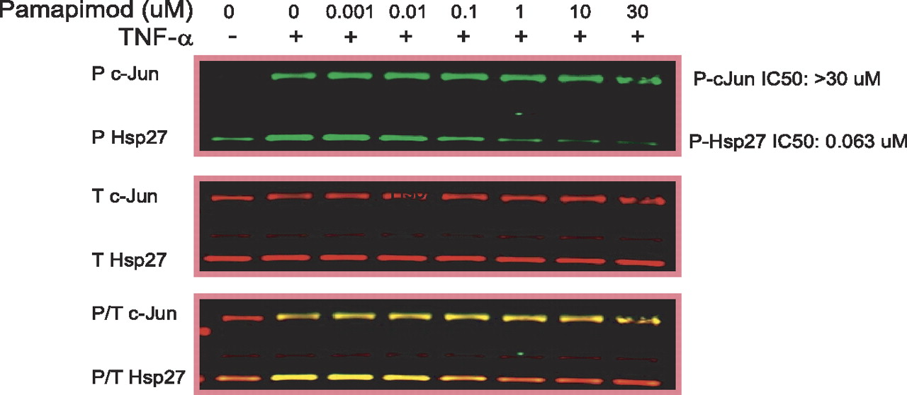
Cellular selectivity of pamapimod for p38 versus Jnk in chondrosarcoma cells. SW-1353 chondrocytes were stimulated with TNF and analyzed by Western blot as described under Materials and Methods. JNK activity was measured by phosphorylation of c-Jun, and p38 activity was measured by phosphorylation of HSP-27. Green (top), phosphospecific antibodies; red (middle), total protein. The third panel shows an overlay of two colors to represent relative contribution of phosphorylated and total protein. Pamapimod had no effect on Jnk activity but inhibited p38, with an IC50 of 0.063 μM.
Pamapimod Suppresses Spontaneous Production of TNFα by Synovial Explants from RA Patients. To evaluate the potential effects of pamapimod on inflammation in diseased joints of human RA, spontaneous production of TNFα from synovial explants was measured after treatment with pamapimod for 2 days at varying concentrations. As shown in Fig. 5A, pamapimod inhibited TNFα production by the synovial explants with an IC50 (95% confidence interval) of 0.104 (0.048–0.23) μM, which is similar efficacy to that observed for inhibition of LPS-induced cytokine production in human whole blood. Thus, assuming the tissue levels of pamapimod are similar to those achieved in the plasma, the inhibition of cytokine production in whole blood could be a surrogate for inhibition of inflammatory cytokines in the diseased joint and could be used as a pharmacodynamic assay to predict efficacious dosing. In addition, upon comparing the efficacy of pamapimod with that of SB203580 (Fig. 5B), which is the widely used reference compound for p38 inhibition, SB203580 had a similar IC50 (95% confidence interval) of 0.240 (0.015–3.7) μM in the explant model. In performing this study, we observed that there was a subset of patients who were partial responders, i.e., they did not achieve full inhibition with drug. This partial response occurred at a rate of approximately one in three donors.
Efficacy of p38 Inhibition in a Spontaneous Mouse Model of Lupus. To assess the role of p38 in lupus, spontaneous lupus-prone MRL/lpr mice were treated with R9111, an analog of pamapimod with equivalent kinase selectivity (see Supplemental Fig. 2) and potency against LPS-induced cytokine production (TNFα from THP-1 cells, EC50 = 0.03 ± 0.02 μM; IL-1β from whole blood, EC50 = 0.07 ± 0.04 μM). R9111 was administered by minipump beginning at 12 to 14 weeks of age for 2 weeks (Fig. 6). Although vehicle-treated animals exhibited renal disease as evidenced by significant proteinuria and histopathological evidence of lupus glomerulonephritis, perivascular leukocyte infiltration, and occasionally arteritis, treatment with R9111 significantly reduced both proteinuria and total perivasculitis scores (Fig. 6, A and B), the former comparable if not superior to dexamethasone treatment. These findings correlated with the ability of R9111 to inhibit p38 activity in vivo, as evidenced by blockade of phosphorylation of the p38 substrate Hsp25 (Fig. 6C). Thus, in a short-term administration regimen, the analog of pamapimod, R9111, was effective in the treatment of MRL/lpr renal disease.
Discussion
The current study characterized the in vitro and in vivo potency of the small-molecule inhibitor of p38 mitogen-activated protein kinase, pamapimod. A binding assay to profile pamapimod against 350 other protein kinases demonstrated that the molecule is highly selective, binding only to JNK, another MAPK family member, with affinity comparable with p38. However, despite the apparent affinity for JNK, evaluation of Jnk inhibition in two cell types based on phosphorylation of c-Jun demonstrates that in cells, pamapimod is not potent against JNK. Thus, in vivo pamapimod is anticipated to have little or no direct impact on Jnk activity.
The isoform selectivity of pamapimod is similar to that observed for other small-molecule inhibitors of p38 because it only inhibits only the α and β isoforms but not the γ and δ isoforms (Kumar et al., 2003; Saklatvala, 2004). This selectivity is consistent with sequence analysis that indicates the α and β isoforms form a subgroup distinct from that of γ and δ, with only 60% homology between the subgroups (Cohen, 1997). The β isoform is minimally expressed in inflammatory cells and preferentially expressed in endothelial cells (Hale et al., 1999). Data from this study indicate that pamapimod is selective for the α isoform by 34- or 92-fold based on enzyme IC50 or binding Kd, respectively. The δ isoform is expressed by monocytes, macrophages, lymphocytes, and neutrophils (Hale et al., 1999) but is not targeted by pamapimod and, therefore, is not part of the mechanism of action. In accordance, these findings suggest that pamapimod inhibits inflammation through its activity against p38α.
The finding that p38 was a target of the pyridinyl imidazoles that block LPS-induced TNF-α and IL-1β release from monocytes (Lee et al., 1994) initiated over a decade of work to define the role of p38 in inflammatory processes. p38 is ideally positioned to integrate signaling from cytokines and growth factors and to convert these signals into transcription of inflammatory cytokines, matrixmetalloproteinases, and Cox-2 and other effectors; therefore, it has become an attractive target for therapeutic inhibition of RA and other autoimmune diseases (for review, see Kumar et al., 2003; Goldstein and Gabriel, 2005; Schieven, 2005; Westra and Limburg, 2006). p38 regulates cellular responses at multiple levels including transcriptional activation (Han et al., 1997; Wesselborg et al., 1997; Zhu and Lobie, 2000), post-transcriptional mRNA stability (Winzen et al., 1999; Frevel et al., 2003; Dean et al., 2004), translation (Brook et al., 2000), and phosphorylation of downstream kinases such as MAP-KAPK-2 (Brook et al., 2000). In addition, p38 appears to regulate cytokine and chemokine gene expression by recruitment of nuclear factor-κB to cryptic binding sites through a mechanism involving phosphorylation of histone H3 (Saccani et al., 2002). The current study demonstrates that pamapimod inhibits LPS-induced release of TNFα and IL-1β both in vitro and in vivo, which is consistent with the reported role for p38 in regulation of inflammatory cytokines. It also demonstrates that pamapimod inhibits phosphorylation of HSP-27, a substrate of MAPKAPK-2, which indicates p38 signals through MAPKAPK-2.

Pamapimod inhibits clinical disease scores in murine collagen-induced arthritis. Mice were immunized on day 0 with type II collagen and given an i.p. injection of LPS 4 weeks later to accelerate and synchronize disease onset. After detection of disease as assessed by clinical score, mice were treated for 10 days with pamapimod or dexamethasone (Dex). Pamapimod inhibited disease scores in a dose-dependent manner. Both 100 and 150 mg/kg doses produced a statistically significant suppression of disease (*, p < 0.05; **, p < 0.01). The presented data are from a single study representative of three independent experiments.
The effects of pamapimod on collagen-induced arthritis in mice are consistent with previous studies of p38 inhibitors (Badger et al., 1996; Nishikawa et al., 2003; Wada et al., 2005; Medicherla et al., 2006). However, the current study is the first to characterize an exquisitely selective p38 inhibitor, thus strongly suggesting that the observed disease modification occurs solely through a p38 mechanism. This finding is important because collagen-induced arthritis in mice can be inhibited by a variety of immune modulators. In addition, previous compounds shown to have efficacy in the rodent models of arthritis had significant off-target activity that could have contributed to the disease modification and adverse side effects (Fabian et al., 2005; Goldstein and Gabriel, 2005; Zhang et al., 2007). However, it is noteworthy that in a recent phase II trial of pamapimod, elevation of liver transamidase levels and Cmax-related dizziness in some patients was reported (Cohen et al., 2008). Therefore, p38 target selectivity does not eliminate all liver and central nervous system side effects.
The inhibition of bone loss, as demonstrated in this study, also has been reported previously with p38 inhibitors (Badger et al., 1996; Nishikawa et al., 2003; Medicherla et al., 2006). In one other study that reported decreased bone loss with a p38 inhibitor, there was a concomitant decrease in osteoclasts (Medicherla et al., 2006), presumably because of the role of p38 in osteoclast differentiation (Li et al., 2002; Pargellis and Regan, 2003). This outcome is potentially beneficial in treatment of RA with p38 inhibitors because current therapies have poor efficacy against cartilage and bone loss. In addition, the effect of pamapimod on inflammatory pain is another potential benefit in a therapeutic setting. A previous study with a p38 inhibitor, SB203580, also demonstrated reversal of inflammatory mechanical hyperalgesia induced by injection of complete Freund's adjuvant into the hindpaw (Ganju et al., 2001). In addition, there are many studies showing that p38 inhibitors can attenuate inflammatory pain (for review, see Ji et al., 2007).
The elevation of cytokines in rheumatoid synovium is clearly a key component to the ongoing chronic inflammation of RA. Thus, the finding that p38 inhibition suppresses the spontaneous production of TNFα in synovial explants is encouraging because it suggests that pamapimod may inhibit cytokine release in the joints of RA patients. A previous study with the p38 inhibitor SB203580 reported low potency against TNFα produced by rheumatoid synovial explants compared with primary macrophages (Campbell et al., 2004). In this study, however, both pamapimod and SB203580 were effective against TNFα produced by diseased synovial explants as LPS-induced TNFα from other primary human cells. The reason for this discrepancy with previous work is not clear. Our observation that one of three donors only responded partially to inhibition by pamapimod may have significance in predicting clinical response rates.
The involvement of p38 in the response to and production of multiple inflammatory cytokines suggests that inhibition of this target should have utility in other autoimmune diseases in addition to RA. This study demonstrated activity of a pamapimod analog against the spontaneous development of nephritis in MRL/lpr mice, suggesting that p38 inhibition may be an efficacious treatment for lupus as well. A previous study using the same murine animal model implicated p38 in the progression of lupus-like disease by demonstrating elevation of p38 phosphorylation (Iwata et al., 2003). The study also used FR167653, a p38 inhibitor, to reduce kidney pathology, which correlated with a reduction in p38 phosphorylation. Although the compound is reported to be selective, its effects were presumably upstream in the pathway because p38 phosphorylation is not dependent on p38, but rather on the activity of MKK3 and MKK6. The current study demonstrated that direct inhibition of p38 activity as confirmed by a decrease in phosphorylation of a p38-dependent substrate, HSP-25, correlated with reduced proteinuria and histopathology, namely the findings of mononuclear perivasculitis. These data, combined with our selectivity profiling for pamapimod, demonstrate that the amelioration of pathology is dependent on inhibition of p38 activity.

Bone protection by pamapimod in murine collagen-induced arthritis. Micro-CT analysis was used to assess the effects of pamapimod on bone density in the murine collagen-induced arthritis model. A, combined phalanges scores on day 15 post-LPS boost of animals that received 3 to 150 mg/kg pamapimod. Dose-dependent restoration in bone density were observed, reaching significant difference from the vehicle-treated controls at 90 and 150 mg/kg (*, p < 0.05). Dose of 0 mg/kg represents vehicle control. N, naive control. B, representative micro-CT images of paws from controls and 150 mg/kg dosed animals. C, total histology scores corresponding to doses in A. Scoring was performed for cartilage degeneration, periosteal new bone, bone resorption, soft tissue proliferation and inflammation, and joint space pannus and exudates for all animals in a single collagen-induced arthritis study. Total histology scores were then calculated. Statistical analysis was by Wilcoxon rank-sum exact tests. Dose-dependent response is again evident, with significant changes from control achieved at the two highest doses.

Pamapimod inhibits spontaneous TNFα production in RA synovial explants. A, dissociated human RA synovial membrane mixed cell populations were incubated with pamapimod for 2 days, then levels of TNFα in the culture supernatant were determined by ELISA, and the percentage inhibition was calculated. Data are triplicates pooled from eight donors. Spontaneous TNFα levels range from 170 to 2630 pg/ml (n = 8, mean = 947 pg/ml, S.D. = 851). B, identical culture system using SB203580 instead of pamapimod. Data are triplicates from three donors.
In summary, the current study demonstrates pamapimod to be a highly selective inhibitor of p38 when profiled across 350 kinases by competitive binding assays and also when profiled in cells by phosphorylation of selective substrates. This selectivity may translate into a better safety and tolerability profile that could allow full exploration of its pharmacology in humans. The anti-inflammatory activities of pamapimod in cells and in vivo indicate the potential to alleviate the signs and symptoms of RA and to provide bone protection. In addition, the efficacy of a pamapimod analog in an animal model of renal lupus suggests the potential utility of p38 inhibition for autoimmune diseases other that RA.

Efficacy of R9111 in MRL/lpr mouse lupus. Spontaneously lupus-prone MRL/lpr mice were treated by minipump with the p38 inhibitor R9111 or dexamethasone (Dex) at the doses indicated for 2-week duration beginning at 12 to 14 weeks of age. A, renal perivasculitis scores reflected the sum of perivascular scores as assessed in the papillary, arcuate, and perirenal regions on histopathology. For statistical analysis, data were transformed to the ranks of the data, and one-way analysis of variance was applied (**, p < 0.01). B, proteinuria was assessed via determination of protein/creatinine ratio in urine. Log transformation was applied to assure normality, and one-way analysis of variance was applied (**, p < 0.01). C, whole lungs of animals (three animals for each treatment group) were assessed by Western blot for phospho-Hsp25, phospho-p38, total Hsp25, and total p38.
Footnotes
-
Article, publication date, and citation information can be found at http://jpet.aspetjournals.org.
-
doi:10.1124/jpet.108.139006.
-
ABBREVIATIONS: RA, rheumatoid arthritis; MAPK, mitogen-activated protein kinase; TNF, tumor necrosis factor; IL, interleukin; SB203580, 4-(4-fluorophenyl)-2-(4-methylsulfinylphenyl)-5-(4-pyridyl)1H-imidazole; SB202190, 4-(4-fluorophenyl)-2-(4-hydroxyphenyl)-5-(4-pyridyl)-1H-imidazole; LPS, lipopolysaccharide; ELISA, enzyme-linked immunosorbent assay; JNK, c-Jun NH2-terminal kinase; HSP, heat shock protein; TBST, Tris-buffered saline/Tween 20; CT, computerized tomography; HWB, human whole blood; MAPKAPK, mitogen-activated protein kinase-activated protein kinase.
-
↵
 The online version of this article (available at http://jpet.aspetjournals.org) contains supplemental material.
 - Received March 14, 2008.
- Accepted September 4, 2008.

- The American Society for Pharmacology and Experimental Therapeutics


