


Abstract
Interferon-α (IFN-α) is indicated for the treatment of certain viral infections including hepatitis B and C, and cancers such as melanoma. The short circulating half-life of unmodified IFN-α makes frequent dosing (daily or three times weekly) over an extended period (6–12 months or more) necessary. To improve the pharmacokinetics of IFN-α and decrease dosing frequency, IFN-α was fused to human serum albumin producing a new protein, Albuferon. In vitro comparisons of Albuferon and IFN-α showed similar antiviral and antiproliferative activities, although Albuferon was less potent on a molar basis than IFN-α. Pharmacokinetic and pharmacodynamic properties of the fusion protein were enhanced in monkeys. After a single intravenous injection (30 μg/kg,) clearance was 0.9 ml/h/kg, and the terminal half-life was 68 h. After 30 μg/kg subcutaneous injection, apparent clearance (clearance divided by bioavailability) was 1.4 ml/h/kg, the terminal half-life was 93 h, and bioavailability was 64%. The rate of clearance of Albuferon was approximately 140-fold slower, and the half-life 18-fold longer, than for IFN-α given by the subcutaneous route in other monkey studies. Sera from Albuferon-treated monkeys demonstrated dose-related antiviral activity for ≥8 days based on an in vitro bioassay, whereas antiviral activity from IFN-α-treated animals was only slightly elevated relative to vehicle on day 0. Significant increases in 2′,5′-oligoadenylate synthetase mRNA relative to IFN-α- or vehicle-treated animals were maintained for ≥10 days after subcutaneous dosing. The improved pharmacokinetics of Albuferon are accompanied by an improved pharmacodynamic response suggesting that Albuferon may offer the benefits of less frequent dosing and a potentially improved efficacy profile compared with IFN-α.
Interferons (IFNs) are a class of cytokines that play a key role in the regulation of cell growth and differentiation via activation of a cascade of intracellular pathways. Interferons possess antiviral, immunomodulatory, and antiproliferative effects. There are two types of IFNs. Type I includes IFN-α, IFN-β, IFN1Ω, and IFN-κ; type II IFN includes IFN-γ (Foster, 1997; Maeyer and Maeyer-Guignard, 1998;LaFleur et al., 2001). Most cells in the human body produce type I IFNs within hours of viral infection, with the IFN-α family having greater antiviral activity against hepatitis C infection than the others (Hu et al., 2001).
Treatment with unmodified IFN-α for chronic hepatitis C requires frequent injections (e.g., once daily or three times weekly) over the course of therapy due to the molecule's short circulating half-life of 2 to 3 h in humans (Intron A package insert). Although studies have demonstrated the benefit of IFN-α in the treatment of chronic hepatitis C, the regimen of 3 million international units three times per week for 6 to 12 months is effective in less than half of all patients (Thevenot et al., 2001). Among those who do respond to therapy, 50 to 80% relapse within 6 months after dose reduction or treatment discontinuation (Davis et al., 1989; Tine et al., 1991; Fried and Hoofnagle, 1995). Although prolonged therapy (up to 36 months) may improve the sustained response rate (Ahmed and Keeffe, 1999; Damen et al., 2001), it may also lead to an increase in the frequency and severity of adverse events (Medical Letter, 1997). Patient compliance in these long-term dosing regimens is difficult to maintain. The development of more slowly cleared pegylated IFNs has led to reduced dosing frequency and improved responses. The conjugation of polyethylene glycol (PEG) to IFN-α significantly decreases plasma drug clearance, so that treatment using a once-weekly injection schedule is possible. Two versions of pegylated IFN-α are commercially available. PEG Intron (Schering Plough, Kenilworth, NJ), with a linear 12-kDa PEG molecule, has at1/2 in humans of approximately 35 h (Glue et al., 2000). A larger molecule, approved only in Europe at this time, contains a branched 40-kDa PEG molecule and has at1/2 of 77 h in humans (Perry and Jarvis, 2001). In this paper we present an alternative strategy to pegylation, which also permits less frequent dosing. Allometric scaling of pharmacokinetic data from monkeys to humans predicts a longer half-life than that resulting from pegylation of IFN. This might reduce the occurrence of adverse events and increase patient compliance, which has the potential to further increase efficacy.
In an attempt to improve the pharmacokinetic and pharmacodynamic profile of IFN-α, a novel genetic fusion product composed of recombinant human serum albumin and recombinant IFN-α, Albuferon fusion protein (Human Genome Sciences Inc.), has been developed. Albumin is an ideal candidate for conjugated IFN sustained-activity agents as it is the most prevalent naturally occurring blood protein in the human circulatory system, and it has a long half-life of 19 days (Peters, 1996). Albumin has little enzymatic or immunological function and is widely distributed in vivo (Peters, 1996). More importantly, research has shown that therapeutic proteins genetically fused with albumin have longer circulating half-lives and improved stability characteristics (Yeh et al., 1992; Syed et al., 1997).
Albuferon was designed to combine the activity of IFN-α with the pharmacokinetic advantages of a protein such as albumin. The objective is to reduce the dosing frequency while potentially improving efficacy and reducing side effects of conventional IFN therapies. To evaluate the in vitro properties, the antiviral and antiproliferative activities of Albuferon and IFN-α were compared in a number of cell lines. To evaluate the in vivo properties of such a fusion protein, a pharmacokinetic and pharmacodynamic study of Albuferon was conducted in nonhuman primates. The objectives were: 1) to describe the pharmacokinetic behavior of Albuferon after a single intravenous or subcutaneous dose, 2) to evaluate the ability of Albuferon and IFN-α in serum sampled at various time points after drug administration to stimulate antiviral activity in an in vitro bioassay, 3) to evaluate whole blood samples for activation of 2′,5′-oligoadenylate synthetase (2′,5′-OAS) genes after Albuferon and IFN-α treatment as a biomarker of antiviral activity, and 4) to evaluate the immunogenicity of Albuferon.
Materials and Methods
Albuferon and IFN-α
Protein Production.
Albuferon is recombinant human interferon-α (IFN-α) genetically fused to recombinant human albumin. The recombinant protein is composed of recombinant human albumin genetically fused at its C terminus to the N terminus of IFN-α. Albuferon has a molecular mass of approximately 85.7 kDa. Albuferon is produced in a genetically modified yeast strain using methods similar to those presented in Yeh et al., (1992). The construction of the expression vector is described in Chinery and Hinchliffe (1989) and in patents EP 286 424 B and US 5,637,504. Albuferon was fermented at 30°C. The protein was purified from the pooled supernatants using a combination of anion, cation, and semiaffinity chromatography steps. N-terminal sequencing of purified material indicated 95% intact protein. There were no detectable truncation or degradation products found in the preparation. The formulated drug was tested for bioburden, endotoxin, and residual yeast DNA before being released for use in this project.
Interferon-α.
IFN-α-2b (Intron-A; Schering Plough Corp., Kenilworth, NJ) was purchased commercially and stored in accordance with the manufacturer's instructions.
Antiproliferative Activity Assay
The Daudi cell line is a human Burkitt's B cell lymphoma line that is highly sensitive to the antiproliferative effects of type I IFNs (Pfeffer et al., 1998). The antiproliferative activity of Albuferon and IFN-α was evaluated using a [3H]thymidine incorporation assay. Daudi cells (American Type Culture Collection, Manassas, VA) were grown in RPMI 1640 supplemented with 10% fetal calf serum. Cells were seeded at 105/200 μl/well in 96-well plates. Albuferon or IFN-α was diluted in medium and added in 50-μl aliquots to the cultures. Cells were incubated for 5 days. Proliferation was quantitated by pulsing the cells during the last 20 h of culture with 0.5 μCi/well of [3H]thymidine (6.7 Ci/mM; PerkinElmer Life Sciences, Boston, MA). Thymidine incorporation was measured by scintillation counting of triplicate wells. Antiproliferative activity, expressed as EC50, was calculated by nonlinear regression using Prism software (version 3.0; GraphPad Software Inc., San Diego, CA).
Antiviral Activity Assays
The antiviral activity of Albuferon was tested on WISH, MDBK, and COS-1 cell lines (American Type Culture Collection) that had been exposed to encephalomyocarditis virus (EMCV) or vesicular stomatitis virus (VSV). The assay was performed as previously described (LaFleur et al., 2001), on the basis of the protocol established by Rubinstein et al. (1981). Briefly, the cells were seeded in flat-bottomed 96-well plates and grown to 95% confluence. Serial dilutions of Albuferon or IFN-α were added to the wells. After 24 h of incubation, optimal concentrations of the viruses were added. After an additional 24-h incubation, cell monolayers were stained with 1% crystal violet in 15% ethanol. Scoring was accomplished by extraction of stained cells with 70% ethanol/1% acetic acid and absorbance determination at 580 nm in an ELISA microplate reader (Spectra Max 250; Molecular Devices Corp., Sunnyvale, CA). Antiviral activity, expressed as EC50, was calculated by Prism software.
For evaluation of antiviral activity in monkey serum, WISH cells were seeded at 104/well in RPMI 1640 medium/10% fetal bovine serum and allowed to grow to 95% confluence. The medium was removed, fresh medium containing serial dilutions of monkey sera or IFN-α WHO international standard was added to the wells, and 24 h later, the cells were challenged with 2 × 10⁴ plaque-forming units of EMCV. Viable cells were quantified 1 day after virus challenge by dye staining as described above. The titer of the samples was based on the 50% cytopathic effect of the assay. The activity units are expressed as units per milliliter, with the IFN-α serving to generate a standard curve. Data were analyzed by paired two-tailed t test (Prism) comparing antiviral activity in serum from drug-treated animals with that from vehicle controls for each day of the study. Statistical significance was set at P < 0.05.
Drug Administration
Five groups of cynomolgus monkeys, two males and two females per group, were studied as outlined in Table1. Dosing volume for all animals was 0.5 ml/kg. Albuferon was supplied by Human Genome Sciences, Inc. (Rockville, MD), at a concentration of 0.987 mg/ml in a buffered sucrose vehicle solution, which was diluted in vehicle to 0.06 and 0.6 mg/ml for the 30 and 300 μg/kg doses, respectively. IFN-α-2b was purchased commercially, reconstituted with bacteriostatic water for injection on day 0, and stored at 2–8°C for the duration of dosing. The dosing regimen for IFN-α administration was selected on the basis of the schedule of drug administration used clinically for hepatitis C virus treatment.
Experimental design for the single-dose pharmacokinetic and pharmacodynamic study
Blood samples (approximately 1 ml each) were collected for ELISA analyses (pharmacokinetic and immunogenicity assays) via a femoral vein into tubes containing potassium EDTA anticoagulant prior to dosing (0 h) and at the following times after dosing (for Albuferon-treated and vehicle-treated monkeys only): 5, 15, 30, and 45 min; and 1, 2, 4, 6, 8, 12, 24, 48, 144, 192, 240, 288, and 336 h. Samples were stored on wet ice for up to 1 h, centrifuged for plasma harvest, and stored at approximately −20°C. For antiviral activity analysis, blood samples (approximately 0.5 ml) were collected via a femoral vein into tubes without anticoagulant at 0 h and at 24, 48, 96, 192, 240, and 336 h postdose. Samples were allowed to clot at room temperature for up to 1 h, centrifuged for serum harvest, and stored at approximately −20°C. Additional blood samples (approximately 0.5 ml) for antiviral activity and 2′,5′-OAS mRNA analysis were collected via a femoral vein at 0 h and at 24, 48, 96, 192, 240, and 336 h postdose. Samples were placed on ice, treated with TRIzol (Invitrogen Inc.), and stored on dry ice prior to RNA extraction and analysis.
Pharmacokinetic Analyses
Pharmacokinetic samples were analyzed using a commercially available ELISA kit from PBL Biomedical Laboratories (New Brunswick, NJ; kit number 41100), modified to use an Albuferon reference standard for the standard curve and positive controls. A standard curve was prepared from 31.25 to 2500 pg/ml Albuferon in the sample diluent provided. Plasma samples and positive controls were added to a 96-well microtiter plate. The plate was incubated for 1 h at room temperature and then washed. Antibody to IFN-α was added and the plate incubated for 1 h at room temperature. The plate was washed, horseradish peroxidase-labeled antibody to the primary antibody was added, and the plate was incubated for 1 h at room temperature. The plate was washed and tetramethylbenzidine was added. Plates were read at 450 nm using a Spectromax ELISA plate reader (Molecular Devices). The limit of detection was 100 pg/ml. The level of IFN-α in plasma was read using a standard curve for Albuferon and was reported in nanograms per milliliter.
Plasma concentrations of Albuferon after intravenous and subcutaneous administration were analyzed with the software package WinNonlin (version 3.1; Pharsight Corp., Mountain View, CA) using noncompartmental analyses. Standard pharmacokinetic parameters, including clearance (CL or CL/F), volume of distribution (Vz orVz/F), half-life (t1/2), area under the plasma concentration versus time curve (AUC), maximal observed plasma concentration (Cmax), and time to maximal observed plasma concentration (Tmax), were calculated. Plasma concentration data were uniformly weighted for these analyses, and the AUC after subcutaneous dosing was calculated using the linear-up/log-down trapezoidal method. For purposes of calculating AUC0−∞ and clearance, a terminal rate was determined using the slope up to 192 h (prior to the development of anti-Albuferon antibodies).
Plasma concentration profiles for each monkey were analyzed individually, and mean (±S.E.M.) values for the pharmacokinetic parameters are reported. A compartmental analysis was conducted on the data from monkeys administered 30 and 300 μg/kg Albuferon subcutaneously, so that the absorption half-life after subcutaneous dosing could be calculated. The data after subcutaneous dosing were fit to a one-compartment model using first-order input and output. Data were weighted as 1/predicted concentration2(1/Cpred2) for this analysis.
Immunogenicity Analysis
The development of antibodies after IFN-α injection was expected on the basis of reports by Trown et al. (1986) and Bailon et al. (2001). Therefore, in this study, serum samples were assessed for the development of anti-Albuferon antibodies using an ELISA method. Serial dilutions of monkey plasma were added to Albuferon-coated microtiter plates. This was followed by a peroxidase anti-human IgG, A, and M conjugate (Jackson Immunoresearch Laboratory, Inc., West Grove, PA). Tetramethylbenzidine in hydrogen peroxide buffer was used for detection, and absorbance was read at 450 nm. A 2-fold increase in the A450 signal was considered a positive result.
2′,5′-OAS mRNA Quantitation
Total RNA was extracted by TRIzol extraction from blood samples, and 2′,5′-OAS mRNA levels were determined by real-time quantitative PCR using an ABI 7900 Taqman Sequence Detector (Applied Biosystems, Foster City, CA). OAS 1(p42) and OAS 2(p69) mRNA was measured by a one-step reverse transcriptase-polymerase chain reaction (RT-PCR) procedure using the comparative Delta Ct method [Perkin-Elmer (1997) user bulletin no. 2] with 18S ribosomal RNA probe as endogenous reference (Applied Biosystems). The assay was performed using triplicate samples, and the data were normalized by expressing the ratio of 2′,5′-OAS mRNA relative to 18S RNA. Since the data were collected over a time course from each monkey, statistical analysis used repeated measures analysis of variance with autoregressive covariance structure that was heteroscedastic across treatment groups. The covariate structure was selected based upon observation of the data (Naik and Khattree, 1999). Statistical significance was set at P < 0.05.
Human peripheral blood mononuclear cells (PBMCs) were purified from leukapheresis preparations (BRT Laboratories, Inc., Baltimore, MD) by Histopaque (Sigma-Aldrich, St. Louis, MO) gradients. Cells were treated with increasing concentrations of Albuferon or IFN-α for 1, 6, or 20 h, and total RNA was extracted by TRIzol extraction. Induction of mRNA expression was measured by real-time quantitative RT-PCR as described above. Increases greater than 3-fold over baseline were considered to be meaningful.
Results
In Vitro Activity of Albuferon.
The fusion protein was tested for its ability to inhibit the cytopathic effect of viral infection in human (WISH), bovine (MDBK), and simian (COS-1) cell lines. The cells were treated with increasing concentrations of Albuferon or IFN-α and, after 24 h of incubation, infected with EMCV or with VSV. As shown in the representative experiments reported in Fig.1, Albuferon had considerable activity on all the cell lines, with a calculated EC50 of 1.6 ng/ml in WISH cells, 0.12 ng/ml in MDBK cells, and 1.85 ng/ml in COS-1 cells. The results obtained with Albuferon were similar to those reported for various forms of recombinant IFN-α (Kramer et al., 1983;Runkel et al., 1998). Albuferon was most potent on MDBK cells, with a mean EC50 of 0.15 ± 0.05 ng/ml obtained from six independent experiments. In this experimental system, side-by-side comparison of the fusion protein with IFN-α showed that Albuferon is approximately 20 times less potent than IFN-α on a molar basis.

Antiviral activity of Albuferon. The protective effect of Albuferon (closed symbols) and IFN-α (open symbols) at the concentrations indicated was evaluated in antiviral assays using bovine MDBK and simian COS-1 cells challenged with VSV cells or human WISH cells challenged with EMCV as described under Materials and Methods.
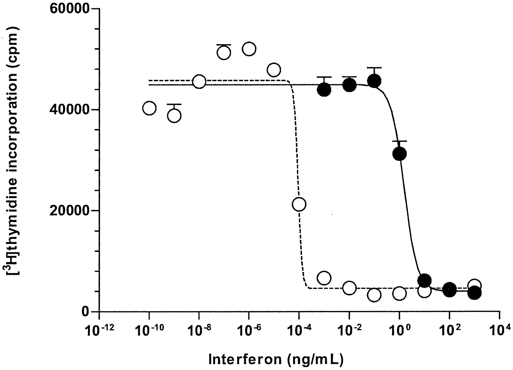
Since the IFNs are multifunctional proteins, we also examined Albuferon effects on the inhibition of cell proliferation and on the regulation of genes relevant for immune-mediated activity. Albuferon strongly inhibited Daudi cell proliferation, as measured by [3H]thymidine incorporation, with an EC50 of 1.52 ng/ml in the experiment shown in Fig. 2. A mean EC50of 2.72 ± 0.72 ng/ml was obtained from four independent experiments.

Antiproliferative activity of Albuferon. Daudi cells were incubated for 5 days in the absence or presence of increasing concentrations of Albuferon (closed circles) and IFN-α (open circles). Cell proliferation was assessed by labeling the cultures with [3H]thymidine during the last 20 h of incubation.
As a further measurement of the biological properties of Albuferon, RT-PCR was used to access the ability of Albuferon to induce mRNA expression of interferon- inducible genes in PBMCs.
PBMCs were treated with 1 and 10 ng/ml of either Albuferon or IFN-α. RNA was extracted and the level of induction was measured relative to untreated cells by quantitative PCR. The average induction levels for a number of target genes are listed in Table2. The numbers represent the mean of two donors. The overall induction of mRNA expression by Albuferon and IFN-α appears to be approximately equivalent at the doses applied for the two donors. Although IFN-α seems to result in a somewhat higher induction for certain target genes, this experiment did not reveal any appreciable differences in potency between Albuferon and IFN-α after taking into consideration the difference in molecular weight between the two molecules.
Induction of mRNA expression by Albuferon and IFN-α in PBMCs
Pharmacokinetics of Albuferon.
Mean plasma concentrations (±S.E.M.) of Albuferon after single intravenous or subcutaneous doses of 30 or 300 μg/kg are shown in Fig. 3. Drug was detectable in all animals through 240 h and in 10 of 12 animals for the 336-h (14-day) duration of the study.

Mean (±S.E.M.) plasma drug concentrations after single-dose intravenous or subcutaneous administration of Albuferon in monkeys (n = 4 per dose/route). Mean plasma concentrations from day 1 to 15 of the study are plotted in the main graph. Plasma concentrations from the first 24 h after dosing are plotted in the inset.
Results of the noncompartmental pharmacokinetic analysis are provided in Table 3. Subcutaneous bioavailability (F) after a dose of 30 μg/kg was approximately 64% relative to the intravenous dose of 30 μg/kg. The absorption of Albuferon after subcutaneous dosing was relatively slow, with an absorption half-life of 25 h for the 30 μg/kg dose and 12 h for the 300 μg/kg dose. Clearance was 0.90 ml/h/kg after the intravenous dose of 30 μg/kg. CL/F was 1.44 ml/h/kg after the subcutaneous dose of 30 μg/kg and 0.94 ml/h/kg after the subcutaneous dose of 300 μg/kg. A markedly increased clearance of Albuferon was observed beginning approximately 10 days after injection. This timing is in good agreement with the timing for the emergence of anti-Albuferon antibodies in the Albuferon-treated monkeys.
Mean (±S.E.M.) pharmacokinetic parameter values after single-dose intravenous or subcutaneous administration of Albuferon in monkeys
Antiviral Activity.
Sera from monkeys treated with Albuferon demonstrated dose-related antiviral activity in the in vitro bioassay. As shown in Fig. 4, serum samples from monkeys treated subcutaneously with 30 or 300 μg/kg Albuferon had significant antiviral activity against EMCV for at least 8 days after injection. Serum samples from monkeys treated with 30 μg/kg intravenously or subcutaneously had similar antiviral activity. Serum from monkeys administered IFN-α on days 0, 2, and 4 had only slightly elevated antiviral activity on day 1 and thereafter demonstrated antiviral activity that was not different from that of vehicle control animals. Sera from monkeys injected with vehicle control had no detectable antiviral activity in this assay.

Mean (±S.E.M.) antiviral activity values in serum from monkeys after single-dose intravenous or subcutaneous administration of Albuferon or multiple-dose subcutaneous administration of IFN-α (n = 4 per dose/route)
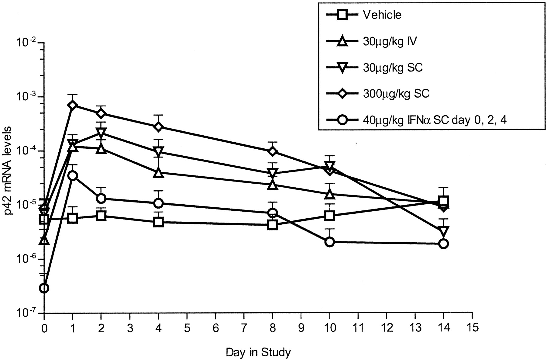
2′,5′-OAS mRNA.
Relative OAS mRNA levels are shown as OAS/18S ratios for 2′,5′-OAS p42 and p69 in Figs.5 and 6, respectively. A single subcutaneous dose of 30 or 300 μg/kg Albuferon resulted in significant increases in 2′,5′-OAS p42 mRNA relative to vehicle controls for up to 240 h after dosing. Monkeys administered a single 30 μg/kg dose intravenously had significant increases relative to vehicle control for 48 h after dosing. Statistically significant changes versus vehicle control were not seen for 2′,5′-OAS p42 mRNA in the animals given IFN-α on days 0, 2, and 4. For Albuferon-treated animals relative to IFN-α-treated animals, significant increases were observed for 30 μg/kg intravenous dose at the 48-h time point; for 30 μg/kg subcutaneous dose at the 48-, 96-, and 240-h time points; and for 300 μg/kg subcutaneous dose from 24 to 240 h after dosing.

Mean (±S.E.M.) 2′,5′-OAS p42 mRNA levels in whole blood after single-dose intravenous or subcutaneous administration of Albuferon or multiple-dose subcutaneous administration of IFN-α (n = 4 per dose/route)

Mean (±S.E.M.) 2′,5′-OAS p69 mRNA levels in whole blood after single-dose intravenous or subcutaneous administration of Albuferon or multiple-dose subcutaneous administration of IFN-α (n = 4 per dose/route)
A single subcutaneous dose of 30 or 300 μg/kg Albuferon resulted in significant increases in 2′,5′-OAS p69 mRNA relative to vehicle controls for 192 h after dosing. Monkeys treated with 30 μg/kg intravenously had significant increases for up to 48 h after dosing. Monkeys treated with IFN-α on days 0, 2, and 4 had significant increases relative to vehicle controls at 24 and 192 h after dosing. For Albuferon-treated animals relative to IFN-α-treated animals, significant differences were observed only for monkeys dosed subcutaneously: 30 μg/kg at the 48- and 96-h time points, and 300 μg/kg from 24 to 96 h after dosing.
Discussion
Albuferon is a fusion product of human albumin and IFN-α and a member of a new class of modified proteins, the human albumin-fusions, which are designed to decrease elimination clearance of otherwise short-acting drugs. This is the first report of preclinical findings based on Albuferon and its comparison to IFN-α. In normal cynomolgus monkeys, the pharmacodynamic activity of Albuferon was shown to be prolonged compared with IFN-α on the basis of its ability to induce elevation of a biological marker of antiviral activity, 2′,5′-OAS mRNA levels. In addition, the results suggest that the enhanced pharmacodynamics of Albuferon are related to its extended circulating half-life, low clearance, and stability in circulation. Albuferon in serum retained antiviral activity for as long as 8 days postinjection in cynomolgus monkeys. Antiviral activity of IFN-α was detected for only 24 h after drug administration. Using a slightly different assay, PEG Intron had detectable antiviral activity for up to 120 h after a subcutaneous dose of up to 14,126 μg/m2 (Food and Drug Administration, 2000).
Albuferon, dosed at 30 or 300 μg/kg subcutaneously or 30 μg/kg intravenously, resulted in detectable plasma concentrations of drug for up to 14 days after dosing. After subcutaneous administration, the fusion product was cleared more slowly (0.9–1.4 ml/h/kg) than the unmodified IFN-α molecule, which was cleared at 214 ml/h/kg (from a previous experiment; data not shown). After subcutaneous administration in monkeys, the half-life of Albuferon was approximately 90 h, whereas the half-life of IFN-α was approximately 5 h (from a previous experiment; data not shown). This is approximately 3 times longer than PEG Intron, which had a terminalt1/2 of 26 h after intravenous injection and 30 to 34 h after subcutaneous injection (Food and Drug Administration, 2000). Subcutaneous bioavailability of Albuferon (64%) is similar to that reported for PEG Intron (57% after a subcutaneous injection of 1413 μg/m2).
In the clinical setting, little or no IFN-α is detected in the patient's blood 24 h after intravenous or subcutaneous dosing (Bailon et al., 2001), and the drug must be dosed once daily or three times a week. Allometric scaling of the clearance based on the work ofMordenti et al. (1991) predicts a half-life of Albuferon in humans in the range of 90 to 288 h and supports the clinical testing of Albuferon administered once every 2 or every 4 weeks. This predicted human half-life is longer than the published human half-lives for the pegylated IFN-α and suggests that Albuferon may be able to be dosed less frequently than either of the pegylated drugs (Glue et al., 2000;Perry and Jarvis, 2001).
IFN-α triggers a series of signal transduction events induced by the binding of IFN-α to its cell surface receptor (reviewed in Foster, 1997; Hu et al., 2001). Downstream events include phosphorylation of Janus kinases and signal transducers and transactivators, formation of IFN-stimulated gene factor 3 and its translocation to the nucleus, and, ultimately, transcription of IFN-sensitive genes that encode IFN-inducible proteins. The enzyme 2′,5′-OAS, in particular, is produced subsequent to IFN-α stimulation. Its antiviral effects are initiated by synthesis of 2′,5′-oligoadenylates that activate an endoribonuclease to cleave double-stranded viral RNA. 2′,5′-OAS is considered to be a reliable biomarker of IFN-α exposure, although its relationship to long-term clinical response is not clear (Moritz et al., 1992; Fischer et al., 1996; Murashima et al., 2000). 2′,5′-OAS mRNA levels were significantly higher after a single Albuferon injection than after three 40 μg/kg IFN-α injections every other day, and the duration of effect was also longer (up to 10 days). Although mRNA induction does not ensure the presence of active protein, it has been reported elsewhere that 2′,5′-OAS activity in the serum of patients treated with IFN-α appears to increase within 6 h of treatment, reaches maximal values at 48 to 72 h, and maintains elevated levels for as long as 4 to 8 months after the initiation of daily IFN treatment (Moritz et al., 1992).
Three different in vitro bioassays of Albuferon have shown that the fusion protein has at least 10-fold lower potency than the parent molecule. However, the enhanced antiviral and OAS activities in vivo indicate that the favorable pharmacokinetic profile of Albuferon compensates for a reduced in vitro potency and contributes to an in vivo pharmacodynamic response that is of greater magnitude and longer duration than the response to IFN-α. In addition, Albuferon is remarkably stable in vivo and retains antiviral activity in monkey serum for up to 8 days after a single injection. In contrast, antiviral activity was only transiently detected in samples obtained from monkeys treated every other day for three injections with IFN-α. Studies of other modified IFN-α molecules (PEG or sulfo-9-fluorenylmethoxycarbonyl groups) have likewise found that in vitro activity is not predictive of biological activity or efficacy in humans (Glue et al., 2000; Motzer et al., 2001; Talpaz et al., 2001).
The emergence of anti-Albuferon antibodies was expected on the basis of published reports of IFN-α administration in monkeys (Trown et al., 1986). In the study reported here, the development of antibodies in 10 of 12 animals coincided with the observed increase in clearance of Albuferon from the circulation. In general, this timing also corresponded with loss of significant antiviral activity and loss of significant levels of 2′,5′-OAS mRNA, at least for subcutaneous dosing. The development of antibodies in these animals was an expected cross-species reaction, since the human Albuferon protein was being administered to a nonhuman species.
Although IFN-α is effective for the treatment of hepatitis C, a disadvantage of IFN-α therapy is that owing to rapid clearance of the drug, treatment must be administered frequently over many months. Pegylated interferons were developed to address this issue. The study reported here presents an alternative strategy, using an albumin-IFN-α fusion protein, that shows reduced clearance of the fusion protein and a longer half-life than has been reported for IFN-α in other cynomolgus monkey studies. The 93-h half-life is 18 times greater than the 5-h half-life determined for IFN-α in cynomolgus monkeys, and 3 times greater than the 30- to 34-h half-life for PEG Intron. Clearance prior to antibody development was 190-fold lower than published pharmacokinetic studies for IFN-α in monkeys (Wills et al., 1984; Collins et al., 1985). Although the advantages of Albuferon are as yet unproven, predicted human half-lives of Albuferon, based on allometric scaling, exceed the published human values for PEG Intron or Pegasys. The reduced dosing frequency may improve patient compliance, improving the response to treatment. In addition, the less frequent dosing may alleviate toxicities associated with peak plasma concentrations by maintaining a more constant circulating drug level with the fusion protein compared with currently available therapy.
The favorable pharmacokinetic and pharmacodynamic properties of Albuferon in this study suggest that this albumin-IFN-α fusion protein may be useful in the clinical setting.
Acknowledgments
We thank Susan M. Stoughton for help in writing this article. The skilled technical assistance of Devanshi Shah, Hsiu Ling Lin, Amal Ahelm, and Nancy Hsu is gratefully acknowledged. We also thank Dr. Deborah Russell for thoughtful reading and review of the manuscript.
Footnotes
-
Financial support was provided by Human Genome Sciences, Inc., Rockville, MD.
-
DOI: 10.1124/jpet.102.037002
- Abbreviations:
- IFN
- interferon
- PEG
- polyethylene glycol
- OAS
- oligoadenylate synthetase
- EMCV
- encephalomyocardititis virus
- VSV
- vesicular stomatitis virus
- ELISA
- enzyme-linked immunosorbent assay
- AUC
- area under the curve
- CL/F
- clearance adjusted for fraction (F) of drug absorbed (bioavailability)
- Cmax
- maximal plasma concentration
- EC50
- concentration to achieve 50% maximal effect
- (bioavailability)
- PBMC, peripheral blood mononuclear cell
- Tmax
- time to maximal plasma concentration
- Vz
- terminal volume of distribution
- Vz/F
- terminal volume of distribution adjusted for bioavailability
- Received May 6, 2002.
- Accepted July 12, 2002.
- The American Society for Pharmacology and Experimental Therapeutics


