


Abstract
Berberine is an isoquinoline alkaloid with multiple pharmacological actions, including anti-inflammatory activity. The aims of this study were to examine the effect of berberine on the mucosal healing process and to investigate whether berberine can inhibit the increased production of interleukin-8 in trinitrobenzene sulfonic acid-induced colitis in rats. Berberine was administered orally for 3 days or 1 week at a dosage of 7.5 or 15 mg/kg/day. Tissue damage scores, body weight, colon wet weight, and colon wall thickness were measured, and myeloperoxidase activity in colon tissue was also examined. Histological lesions, morphological damage, and myeloperoxidase activity were reduced after 1 week of treatment with berberine at a dosage of 15 mg/kg/day. Furthermore, 1 week after trinitrobenzene sulfonic acid treatment, the production of interleukin-8 by cultured rectal mucosa or cardiac blood mononuclear cells with or without stimulation of lipopolysaccharide for 24 h was also analyzed by enzyme-linked immunosorbent assay. Cardiac blood mononuclear cells and rectal mucosa of normal rats produced substantial amounts of interleukin-8, which increased strikingly with the stimulation of lipopolysaccharide. Cardiac blood mononuclear cells and rectal mucosa of trinitrobenzene sulfonic acid-treated rats secreted more interleukin-8 than those of normal rats. The addition of berberine with a concentration of 10−5 M to the culture media resulted in an inhibition of interleukin-8 production of rectal mucosa.
Berberine is the major alkaloid distributed in Goldenseal (Hydrastis canadensis) (Jang, 1941), which is a popular herb in the United States. In Chinese medicine, Coptidis rhizoma andPhellodendri cortex have been used to treat the patients who have gastroenteritis, abdominal pain, or diarrhea. Berberine was also identified as a major component in those plants and proved to have biological activities, such as bacteriocidal activity, an anticholera toxin effect, and an anti-inflammatory effect. Its structural formula is shown in Fig. 1. In this study, we characterized the effect of berberine on trinitrobenzene sulfonic acid (TNB)-induced colitis in rats in vivo and in vitro.
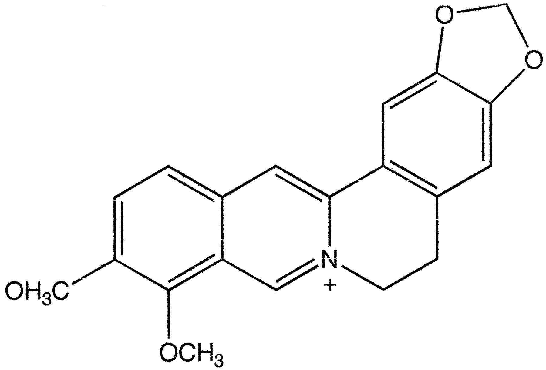
Structural formula of berberine.
The pathological features of inflammatory bowel disease (IBD), such as Crohn's disease and ulcerative colitis, are marked by the presence of mucosal ulceration and the infiltration of neutrophils and lymphocytes in the mucous membranes. Although immunologic mechanisms have been postulated to play an important role in the pathogenesis of these diseases, the etiology remains obscure. The mainstay of medical therapy focuses on inhibition of the effect of the inflammatory mediators operant in IBD because the causes of these two chronic disorders are unknown. To investigate the etiology of IBD and the effects of drugs on tissue damage, animal models have been reported as useful. In particular, TNB-induced colitis has been suggested as the more relevant model of IBD because this model involves the use of an immunologic hapten that results in acute inflammation with ulcers that evolves into chronic inflammation of the distal colon (Morris et al., 1989). Glucocorticoid and salicylazosulfapyridine have been mainly used for the treatment of these diseases, but their side effects remain a major clinical problem. Consequently, traditional Chinese herbal medicine has recently generated increasing interest for the treatment of these disorders. We have reported the beneficial effects of traditional Chinese medicine on refractory steroid-dependent and steroid-resistant ulcerative colitis in human (Zhou et al., 1995) and the beneficial effects of Oren-gedoku-to (a complex mixture of ingredients derived from C. rhizoma, Scutellariae radix, P. cortex, andGardeniae fructus plants) on TNB-induced colitis in rats (Zhou and Mineshita, 1999a). Although the precise mechanism of action for Oren-gedoku-to and the whole constituents of Oren-gedoku-to are still unclear; berberine, the main constituent of C. rhizomaand Phellodendri cortex, may play an important role on the mucosal healing process and reduction of interleukin-8 (IL-8). The enhanced production of potent inflammatory cytokine IL-8 in IBD has been studied (Nielsen et al., 1987; Daig et al., 1996). In experimental studies, it has also been demonstrated that berberine inhibits cyclooxygenase-2 transcriptional activity (Fukuda et al., 1999) and lipoxygenase activity (Misik et al., 1995).
The aims of this study were to investigate the production of IL-8 in normal and inflamed rat colon and to evaluate the effect of berberine on IL-8 production by cultured rectal mucosa or cardiac blood mononuclear cells obtained from rats with colitis induced by TNB in vitro. Furthermore, we examined the ability of berberine to modify the course of mucosal changes in TNB-induced colitis in rats.
Materials and Methods
Animals
Male Sprague-Dawley rats (180–220 g), obtained from Saitama Animal Laboratories (Saitama Prefecture, Japan), were used. They were kept in a restricted-access room with a controlled temperature (22° ± 2°C) and light/dark cycle (12/12 h). Three animals were housed in each standard wire mesh cage, and they were fed standard pellets with tap water freely available. All of the animal studies adhered to the guidelines established by the Tokyo Medical and Dental University for the care and use of laboratory animals.
In Vivo Experiment
Colitis Induction.
Experimental colitis was induced using TNB as described by Morris et al. (1989). In brief, rats that had been fasted for 16 h were lightly anesthetized with pentobarbital sodium (20 mg/kg i.p.), and then 0.25 ml of 50% ethanol containing 30 mg TNB was infused intrarectally via a catheter. The tip of the catheter was inserted after removing distal stools so it was set at 7 cm proximately to the anus. To clear the TNB/ethanol solution from the catheter, 0.5 ml of air was injected, and then the rats were left for 5 min in a supine Trendelenburg position with the anus clipped. Tissue paper was laid in the cages and changed twice a day to minimize the pain and distress of the rats due to diarrhea after TNB treatment for 1 week. The control group of rats were also subjected to the same surgical procedures as described, but they were injected with normal saline instead of TNB.
Experimental Design.
TNB/ethanol colitis was induced in 30 rats, which were then randomized into three groups to receive a daily oral administration of berberine (7.5 mg/kg, n = 10; 15 mg/kg, n = 10) or an equal volume of distilled water (n = 10) from day 1 (24 h after the induction of colitis) for 3 days or 1 week, when the animals were sacrificed. An additional 10 rats, which were used as control animals, received an injection of saline instead of TNB, and distilled water was given from day 1 for 1 week. Observations were made at 3 days and 1 week after the intrarectal administration of TNB. The rats were euthanized by pentobarbital sodium (90 mg/kg i.p.), and a 10-cm segment of the distal colon was removed for the biochemical and morphological studies.
Body Weight Measurement.
The rats were weighed between 9:00 and 10:00 AM on an electric balance every day before and after the TNB treatments for 1 week.
Macroscopic Assessment of Colonic Damage.
On the 3rd day and 1 week after the intrarectal administration of 30 mg of TNB in 50% ethanol, five rats from each group were randomly selected and euthanized by pentobarbital sodium (90 mg/kg i.p.). The distal colons were removed, opened by longitudinal incisions, and assigned code numbers. Each colon was assessed by a semiquantitative scoring system, as described previously (Vilaseca et al., 1990). in a blind trial procedure. Table 1 shows the macroscopic and microscopic scoring system that was used.
Criteria for assessment of colonic damage
Histological Evaluation of Colonic Damage.
After macroscopic scoring, the mass of colon (7 cm from the anus) was taken out and weighed at once. Then two tissue samples (2 × 10 mm) were excised from the central part of the lesion of each colon. When there was no inflammation macroscopically, at least one sample was taken. The tissue samples were fixed in 4% buffered formaldehyde and prepared for routine paraffin embedding. Sections of tissue were cut into 5-μm slices on a microtome, mounted onto slides, and then stained with H&E. Histological assessment by light microscopy was performed in a blind method on coded slides using the criteria of Vilaseca et al. (1990) as outlined in Table 1. As an index of tissue edema, the thickness of the colonic wall was determined by measuring the distance from the serosal surface to the luminal surface of the mucosa. The diameter of ulcers was also measured.
The mucosa, submucosa, and muscularis propria were separately evaluated for infiltration of inflammatory cells, such as neutrophils, eosinophils, macrophages, and lymphocytes. A scale of 0 to 3 was used to semiquantify inflammatory cell infiltration: 0, 0 to 10 leukocytes per high-power field (HPF); 1, 11 to 25 leukocytes/HPF; 2, 26 to 50 leukocytes/HPF; and 3, ≥51 leukocytes/HPF. At least 8 HPFs from each section were examined (Zipser et al., 1987).
Myeloperoxidase (MPO) Activity Measurement.
MPO is an enzyme that is found predominantly in the azurophilic granules of neutrophils and has been used as a quantitative index of inflammation in several tissues, including the intestine (Krawisz et al., 1984). Samples of colon were obtained from the most inflamed part of lesion after the samples for histological examination were derived. Samples were immediately washed in cold PBS, pH 7.4, and blotted to remove the interfering blood and dirt. Subsequent to weighing on an analytical balance, the distal segment of the colon (150–300 mg) was suspended in 1.0 ml of ice-cold 0.5% hexadecyltrimethylammonium bromide in 50 mM phosphate buffer, pH 6.0, and then homogenized three times for 30 s using a tissue homogenizer (OMNI GLH-2017; Yamato Scientific Co., Ltd., Tokyo, Japan). The probe was rinsed twice with 1.0 ml of the buffer. After the homogenate thawed and froze three times, it was centrifuged for 15 min at 12,000g. The tissue level of MPO activity was measured spectrophotometrically: 0.1 ml of supernatant was combined with 2.4 ml of 60 mM phosphate buffer, pH 6.0, containing 0.2 mg/ml O-dianisidine hydrochloride. After preincubation for 10 min at 25°C, 0.5 ml of 0.003% hydrogen peroxide was added to the buffer; after incubation for an additional 10 min, 0.5 ml of 0.1% sodium azide was added to stop the reactions. The absorbance at 460 nm was measured on a spectrophotometer (DU-640; Beckman Instruments, Inc., Fullerton, CA). A unit of MPO activity was defined as that converting 1 μmol of hydrogen peroxide to water in 1 min at 25°C and was divided by colonic wet weight. The MPO assay was performed in a blind fashion on coded tubes.
In Vitro Experiment
Experimental Design.
Cardiac blood mononuclear cells (CBMCs) and rectal mucosa were obtained from six normal rats and six rats with TNB-induced colitis. The cells and rectal mucosa were cultured with or without stimulation of lipopolysaccharide (LPS) derived from theEscherichia coli strain 0111:B4 for 24 h. Berberine at a concentration of 10−6 or 10−5 M was also added to the culture medium, which was stimulated by LPS. IL-8 levels in the medium were measured by enzyme-linked immunosorbent assay method using a commercial kit.
CBMC Culture.
One week after TNB administration, six rats with TNB-induced colitis and six normal rats (control group) were euthanized with pentobarbital sodium (90 mg/kg i.p.). CBMCs were separated as previously described by Boyum (1968) using Ficoll-Paque solution. Cardiac blood diluted with balanced salt solution (1:1) containing 100 I.U. heparin/ml was layered over Ficoll-Paque (Pharmacia Biotech, Uppsala, Sweden) and centrifuged for 40 min at 400g. Cells harvested from the interface were washed three times in Ca2+- and Mg2+-free PBS and resuspended at a final concentration of 2 × 10⁶/ml in a culture medium consisting of RPMI 1640 supplemented by 10% heat-inactivated FBS, 2 mM l-glutamine, 100 I.U./ml penicillin, and 100 μg/ml streptomycin. Thereafter, the rat CBMCs were incubated with doses of berberine ranging from 10−6 to 10−5 M, with or without simultaneous activation by 100 μg/ml LPS in 24-well culture plates (Falcon; Polylabo, Strasbourg, France) for 24 h at 37°C in a humidified 5% CO2, 95% air atmosphere. Supernatants were then removed, filtered (0.45 μm), and stored at −80°C until IL-8 measurement. The cell viability was assessed by the trypan blue exclusion test (Phillips, 1973).
Intestinal Specimens and Tissue Culture.
One week after TNB administration, rats were euthanized as described before. Rectal mucosal specimens were scraped from the marked inflamed part, avoiding ulcerative areas, and placed in Ca2+- and Mg2+-free PBS supplemented with 100 I.U./ml penicillin and 100 μg/ml streptomycin at 4°C for organ culture. After collection, the tissue was gently washed three times in PBS with added penicillin and streptomycin, blotted carefully, weighed (22–25 mg), and placed in 24-well tissue culture plates in 1 ml of RPMI 1640 culture medium with or without simultaneous activation by 100 μg/ml LPS. Berberine (10−6 to 10−5 M) was also added into the culture medium, which was exposed to LPS. After 24 h in culture at 37°C in a humidified 5% CO2, 95% air atmosphere, supernatants were removed, filtered (0.45 μm), and stored at −80°C until IL-8 measurement.
Measurement of IL-8 Production in Culture Medium.
Culture supernatants were diluted 1:2 to 1:30 in assay buffer, and IL-8 levels were quantified with a commercially available rat IL-8 enzyme-linked immunosorbent assay system (Amersham Pharmacia Biotech UK Limited, Amersham, England) according to the manufacturer's instructions. The detection limit of this assay was 4.7 pg/ml. The absorbance at 450 nm was measured on a 96-well plate reader. The levels of IL-8 in culture medium were expressed as picograms of immunoreactive IL-8 per milliliter of medium.
Chemicals and Reagents
For the in vivo experiment, berberine chloride (purity of 99.0%; Sigma Chemical Co., St. Louis, MO) was suspended in distilled water and shaken for 30 min. For the in vitro experiment,berberine chloride (purity of 99.0%; Wako Pure Chemical Industries, Ltd., Osaka, Japan) was suspended in culture medium that adjusted the concentration to 10−6 and 10−5 M.
TNB was also obtained commercially from Sigma Chemical Co. It was dissolved in 50% ethanol and shaken for 30 min before use.
LPS from E. coli 0111:B4 was obtained commercially from Wako Pure Chemical Industries, Ltd. It was dissolved in distilled water and adjusted to the concentration of 100 μg/ml before use.
Hexadecyltrimethylammonium bromide and hydrogen peroxide for MPO activity measurement were obtained commercially from Wako Pure Chemical Industries, Ltd. O-Dianisidine hydrochloride was obtained commercially from Sigma Chemical Co.
For cell and organ culture, RPMI 1640, l-glutamine, fetal beef serum (FBS), and trypan blue were obtained commercially from GIBCO BRL, Life Technologies (Grand Island, NY). Penicillin and streptomycin were obtained commercially from Sigma Chemical Co.
Statistical Analysis
All values are expressed as mean ± S.D. The Student'st test and Mann-Whitney U test were used for parametric and nonparametric statistical evaluation, respectively. A value of P < .05 was considered as the level of significance.
Results
General Observations
Intrarectal administration of TNB/ethanol resulted in an inflammatory response characterized by extensive mucosal disruption, linear and deep ulcers, hemorrhage, and submucosal edema. The adhesions between colon and small bowel and other organs were seen in 90% of the rats. Diarrhea and lack of weight gain were seen in all rats without berberine treatment.
In Vivo Experiment
Effect of Berberine on Body Weight.
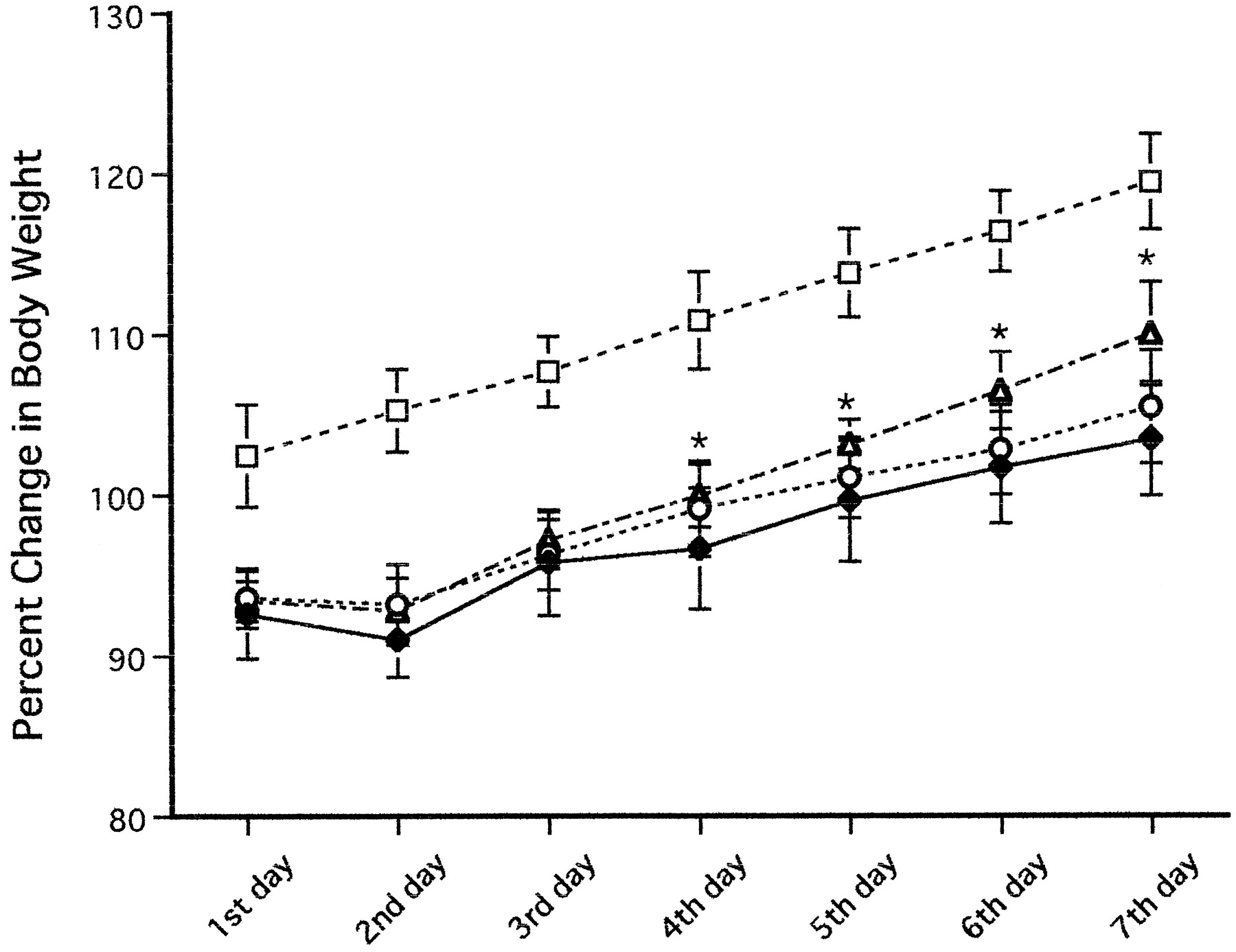
The effect of berberine on body weight change is shown in Fig. 2. Body weight is expressed as the percentage of weight on the day before TNB treatment. The body weight of rats treated with 30 mg of TNB decreased throughout the 5-day post-treatment period and increased slightly on the 7th day. The group given berberine (15 mg/kg) for 1 week showed quicker recovery of their body weight. However, there were no differences between the two groups treated with berberine at the 3rd day and between the group treated with a low dose of berberine at the 7th day and the TNB control group.

Effects of berberine chloride on rat body weight (blank, ■; berberine chloride 7.5 mg/kg, ○; berberine chloride 15 mg/kg, ▵; TNB, ♦) from 1st day to 7th day after administration of 30 mg of TNB. The body weight is expressed as the percentage of weight on the day before TNB treatment. Berberine chloride increased the body weight on 4th to 7th days in the dose of 15 mg/kg. Results are mean ± S.D. (n = 5). *P< .05 compared with the inflamed untreated group.
Effect of Berberine on Macroscopic Damage.
Figure3A shows the macroscopic scores obtained from rats euthanized 3 days and 1 week after the TNB (30 mg) administration. At 1 week, the mean macroscopic damage scores were lower in those rats that had been treated with berberine (15 mg/kg) than in the TNB control group, but there were no significant differences among the other groups. Figure4 shows the macroscopic views of the colons that were obtained from TNB control and berberine-treated rats euthanized at various times later. The development of chronic inflammatory lesions in the colon was mitigated by the berberine at a dosage of 15 mg/kg for 1 week. However, there were no differences in the development of chronic inflammatory lesions between the two berberine groups at the 3rd day and between the group given a lower dose of berberine at 1 week and the TNB control group.

Effects of berberine chloride on macroscopic (A) and microscopic (B) damage score 3 days and 1 week after the induction of colitis. Table 1 shows colonic damage was scored on a scale of 0 to 10 (macroscopically) and 0 to 8 (microscopically) (TNB, ●; berberine chloride 7.5 mg/kg, ○; berberine chloride 15 mg/kg, ⋄). Berberine chloride (15 mg/kg) reduced the colonic damage score (macroscopic or microscopic) at 1 week significantly. Results are mean ± S.D. (n = 5). *P < .05, **P < .01.
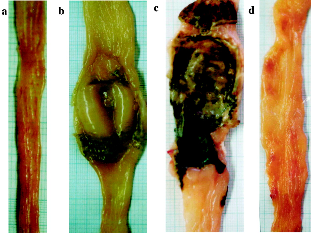
Photographs of colons. a, normal rat. b, rat administered 30 mg of TNB alone and obtained at the 3rd day after administration. Note the extensive hyperemia, edema, and ulceration of the colon. This colon was given a damage score of 10. c, rat administered TNB alone and obtained at 1 week after administration. Note the extensive large ulceration of the colon and the grossly visible enlargement of the colon. This colon was given a damage score of 10. d, rat treated with berberine chloride (7.5 mg/kg) and obtained 3 days later. This colon was given a damage score of 9. e, a rat treated with berberine chloride (15 mg/kg) and obtained 1 week later. This colon was given a damage score of 4.
Effects of Berberine on Histological Damage Score.
The histological appearance of tissues examined at 3 days and 1 week after the administration of TNB (30 mg) showed tissue damage characterized by edema, hemorrhage, epithelial exfoliation, and infiltration of polymorphonuclear leukocytes, macrophages, eosinophils, and lymphocytes. There was more inflammatory cell infiltration at 1 week after TNB administration than that of the 3rd day. As shown in Fig. 3B, the mean histological damage score of the distal colon was lower at 1 week in the group given berberine (15 mg/kg), but there were no significant differences between the two berberine groups at the 3rd day and between the group given low-dose berberine at 1 week and the TNB control group. Figure 5 shows the histological sections of the intestines obtained from the TNB control and berberine-treated rats at 1 week.

Histological appearance of colon from rats with TNB-induced colitis 1 week later (H&E; magnification, 40×; A), showing mucosal ulceration (top right) and transmural inflammation most evident in the enlarged submucosa; and of colon from berberine chloride-treated rat 1 week later (H&E; magnification, 40×; B), showing healing of lesions and restored mucosa.
Effects of Berberine on Wet Weight and Wall Thickness of Colon.
Table 2 shows the mean wet weight of distal colons (7 cm) and the mean intestinal wall thickness at the site of grossly visible ulceration and inflammation at 3 days and 1 week after the colitis was induced by 30 mg of TNB. Wet weight and wall thickness of the colon were decreased at 1 week in the group given berberine (15 mg/kg) compared with the control group. However, there were no differences between the two berberine groups at the 3rd day and between the low-dose berberine group at 1 week and the TNB control group.
Effect of berberine chloride on wet weight of colon and intestinal wall thickness
Effect of Berberine on Ulceration of Colon.
The ulceration of colon was examined at 3 days and 1 week after the colitis was induced by 30 mg of TNB. As shown in Table 3, the mean diameter of the colonic ulcers was smaller at 1 week in the group given berberine (15 mg/kg) than in the control group, but there were no significant differences between the two berberine groups at the 3rd day and between the low-dose berberine group at 1 week and the TNB control group.
Effect of berberine chloride on intestinal ulceration
Effects of Berberine on MPO Activity in Inflamed Colonic Tissues.
As shown in Table 4, MPO activity was significantly increased in TNB animals compared with controls. In the group treated with berberine (15 mg/kg), the MPO activity of colonic tissues was lower at 1 week than in the TNB control group, but there were no significant differences between the two berberine groups at the 3rd day and between the low-dose berberine group at 1 week and TNB control group.
Effect of berberine chloride on MPO activity of intestinal wet tissue
In Vitro Experiment
Macroscopic Damage Score of Colons.
One week after TNB administration, the macroscopic damage score of colons was 8, 9, 9, 8, 10, and 8, respectively, for the six colitis rats and 0, 0, 0, 0, 0, and 0, respectively, for the control rats.
Effects of Berberine on Production of IL-8.
As outlined in Table 5, the CBMCs and rectal mucosa of TNB-induced colitis rats and normal rats produced substantial amounts of IL-8, which increased remarkably when stimulated with LPS (100 μg/ml). Levels of IL-8 secreted by CBMCs or rectal mucosa of TNB-treated rats in the absence or presence of LPS were significantly elevated compared with the control group (P < .01). The IL-8 production in rectal mucosa was inhibited by the addition of berberine into the culture medium with a concentration of 10−5 M but not with a concentration of 10−6 M. However, the IL-8 production of CBMCs was not affected by both concentrations of berberine. The cell viability was not affected by an addition of berberine at both concentrations.
The effect of berberine chloride on IL-8 production in cultured CBMCs and rectal mucosa
Discussion
In the present study, the administration of berberine markedly reduced the inflammation in rats with TNB-induced colitis, as verified by effects on macroscopic and histological damage and MPO activity. The IL-8 production in cultured mucosa of inflamed colon was increased compared with noninflamed colon. The addition of berberine with a concentration of 10−5 M to the culture medium inhibited the IL-8 production in response to LPS in vitro. This suggests that the inhibitory effect of berberine on colonic IL-8 production may contribute to the tissue healing process, although a direct cause-and-effect relationship must be proved.
Our results showed a beneficial effect of berberine on the inflammatory response in TNB-induced colitis in rats in vivo. One week of treatment with berberine reduced the colonic mucosal inflammation as demonstrated by macroscopic and histological examination, analysis on colonic wet weight, and MPO activity. This is in agreement with our previous findings that showed similar anti-inflammatory effects of Oren-gedoku-to on TNB-induced colitis in rats (Zhou and Mineshita, 1999a). Oren-gedoku-to is a traditional Chinese herbal medicine that consists of a mixture of C. rhizoma, S. radix,P. cortex, and G. fructus. Although the entire composition of each of these herbal medicines is not yet completely known, the investigations of the chemical compositions and immunological properties show that their activities are mainly due to the major alkaloid berberine in C. rhizoma and P. cortex, baicalin and baicalein in S. radix, and geniposide in G. fructus. We have previously reported that baicalein inhibits IL-8 production of cultured blood mononuclear cells and rectal mucosa (Zhou and Mineshita, 1999b), and our unpublished data have shown that geniposide has no effect on TNB-induced colitis at an oral administration of 10 to 20 mg/kg/day for 1 week. However, the mechanism of the anti-inflammatory actions of Oren-gedoku-to seems to be more complex. Our results in the present study showed that the major constituent of berberine may play an important role in attenuating the inflammation induced by TNB.
It has been suggested that increased intestinal permeability is a primary etiologic factor in both IBD (Stein et al., 1998) and TNB-induced colitis (Yamada et al., 1992). Furthermore, alterations in active electrolyte transport by the diseased epithelium would result in altered water flux and could thereby contribute to the secretory diarrhea that is a frequent symptom of IBD. It has also been reported that berberine inhibits ion transport in human colonic epithelia and decreases the intestinal permeability (Taylor et al., 1999). Those previous reports can help us to explain in part the effect of berberine on TNB-induced colitis.
IL-8 is an important cytokine for the recruitment and activation of polymorphonuclear neutrophils cells that are abundant in the intestinal lesions of IBD. It has been previously reported that locally generated IL-8 is involved in neutrophil migration and binding to extracellular matrix (Ina et al., 1997). Increased mucosal generation of IL-8 may attract neutrophils from the circulation into the inflammatory site and induce binding of neutrophils in the interstitial tissue, contributing to the accumulation and activation of neutrophils in the affected mucosa with IBD. A large number of studies have shown increased levels of IL-8 in ulcerative colitis and Crohn's disease (Izzo et al., 1992,1993; Mitsuyama et al., 1994; Izutani et al., 1995; Daig et al., 1996). Therefore, the inhibition of the IL-8 production is believed to provide a novel approach to the treatment of IBD.
Berberine is an isoquinoline alkaloid present in numerous plants of the genera Berberis and Coptis. It has a wide range of pharmacological and biological activity, including anti-inflammatory and antimicrobial properties. The anti-inflammatory and immunosuppressive activity of berberine is well established by previous investigators (Akhter et al., 1977; Li et al., 1989; Teh et al., 1990;Kondo et al., 1992; Seow et al., 1992; Ivanovska and Philipov, 1996). Therefore, berberine has been used in traditional Eastern medicine for more than 2 millennia in the treatment of bacterial or secretory diarrhea and gastroenteritis (Lahiri and Dutta, 1967; Tang and Eisenbrand, 1992). Its therapeutic benefits have been attributed in part to antimicrobial (Iwasa et al., 1998) and antisecretory (Tai et al., 1981; Taylor et al., 1999) actions. In addition, the inhibitory effect of berberine on cyclooxygenase-2 has been demonstrated (Fukutake et al., 1998; Fukuda et al., 1999). Miura et al. (1997) reported the inhibitory action of berberine on apoptosis; and Ckless et al. (1995)reported the inhibition of in vitro lymphocyte transformation by berberine. The lipoxygenase inhibition and antioxidant properties of berberine have also been reported by Misik et al. (1995). However, the effect of berberine on IL-8 in IBD has not yet been reported. In the present study, we have shown the beneficial effect of berberine on the mucosa healing process, possibly by inhibition of IL-8 production, in TNB-induced colitis in rats both in vivo and in vitro. These findings may have potentially important implications for the therapeutic use of berberine in IBD, in part by disrupting the cycle of cellular mediators.
Acknowledgments
We thank Prof. K. Ohya, Prof. S. Kasugai, and Dr. H. Kondo from the Department of Dental Pharmacology, Dr. T. Kuroiwa and T. Tajima from the Department of Neuropathology, and Asst. Prof. K. Shiba and K. Sugimoto from the Department of Hygiene, Tokyo Medical and Dental University, for fruitful advice and expert technical assistance. We also thank Dr. Guy D. Eslick, and Dr. Harry H. Xia from the Department of Medicine, University of Sydney, Australia, for valuable advice.
Footnotes
-
Send reprint requests to: Dr. Satoru Mineshita, Department of Preventive Medicine, Medical Research Institute, Tokyo Medical and Dental University, 1-5-45 Yushima Bunkyo-Ku, Tokyo 113-8510, Japan. E-mail: satoru-mineshita.prm{at}mri.tmd.ac.jp
- Abbreviations:
- TNB
- trinitrobenzene sulfonic acid
- IBD
- inflammatory bowel disease
- MPO
- myeloperoxyidase
- LPS
- lipopolysaccharide
- CBMC
- cardiac blood mononuclear cell
- IL
- interleukin
- Received March 6, 2000.
- Accepted May 15, 2000.
- The American Society for Pharmacology and Experimental Therapeutics


