


Article Figures & Data
Figures
- Fig. 2.
Chronic alcohol feeding enhanced PARP expression, pADPr synthesis, and NAD+ depletion in alcohol-fed mice. (A) Total proteins were extracted from liver tissues and subjected to Western blot analysis for expression of PARP, pADPr, SREBP-1c, DGAT1, and DGAT2 with specific antibodies. (B–D) Chronic alcohol exposure increased the expression of hepatic PARP protein (B), which catalyzed pADPr formation (C) and lowered hepatic NAD+ content compared with the PF group (D). (E–G) Densitometric analysis showed that chronic alcohol feeding increased the expression of SREBP-1c (E), DGAT1 (F), and DGAT2 (G) proteins. (H–J) Relative gene expression of SREBP-1c (H), DGAT1 (I), and DGAT2 (J) was significantly higher in mice with chronic alcohol consumption compared with the PF group. Values are means ± S.D. (n = 6 per group). *P < 0.05 (significant differences compared with the PF group).
- Fig. 3.
Inhibition of PARP activity augmented NAD+ levels and decreased intracellular TG content in hepatocytes. HepG2 cells were exposed to complete DMEM containing PJ34 (1 µM) for 24 hours. (A) Expression of intracellular PARP and pADPr was detected by Western blot analysis. (B and C) PJ34 treatment did not significantly change the expression of PARP (B) and decreased the synthesis of pADPr (C). (D) PJ34 treatment elevated intracellular NAD+ content. (E) Moreover, PJ34 treatment also decreased intracellular TG content. Values are means ± S.D. from three or more independent batches of cells. *P < 0.05 (compared with the control group).
- Fig. 4.
Inhibition of PARP activity suppressed the expression of critical genes in TG anabolism. HepG2 cells were treated with PJ34 (1 μM) in the presence of OA for 24 hours. Total RNAs were extracted from cells. The mRNA of critical genes was detected by real-time PCR. (A–C) PJ34 treatment changed little in intracellular SREBP-1c (A) and lowered intracellular gene expression of DGAT1 (B) and DGAT2 (C). Values are means ± S.D. from three or more independent batches of cells. *P < 0.05 (compared with control group).
- Fig. 5.
PJ34 injection attenuated hepatic TG accumulation in alcohol-fed mice. C57BL/6 mice were fed an alcohol-containing liquid diet with or without PJ34 intraperitoneal injection for 4 weeks. (A and B) Compared with the AF group, PJ34 injection augmented body weight (A) and lowered the liver/body weight rate (B). (C–E) However, PJ34 injection did not significantly change the epididymis fat/body weight rate (C) and circulating ALT and AST levels (D and E). (F–I) PJ34 intraperitoneal injection decreased hepatic TG content (F) and circulating TG levels (G) but had no difference on hepatic TC content (H) and the level of circulating TC (I) compared with the AF group. (J) Liver sections were stained with H&E. (K) The quantitative statistics of lipid droplet fold change are shown. Values are means ± S.D. (n = 6 per group). *P < 0.05 (compared with the AF group). Original magnification, ×400 in (J).
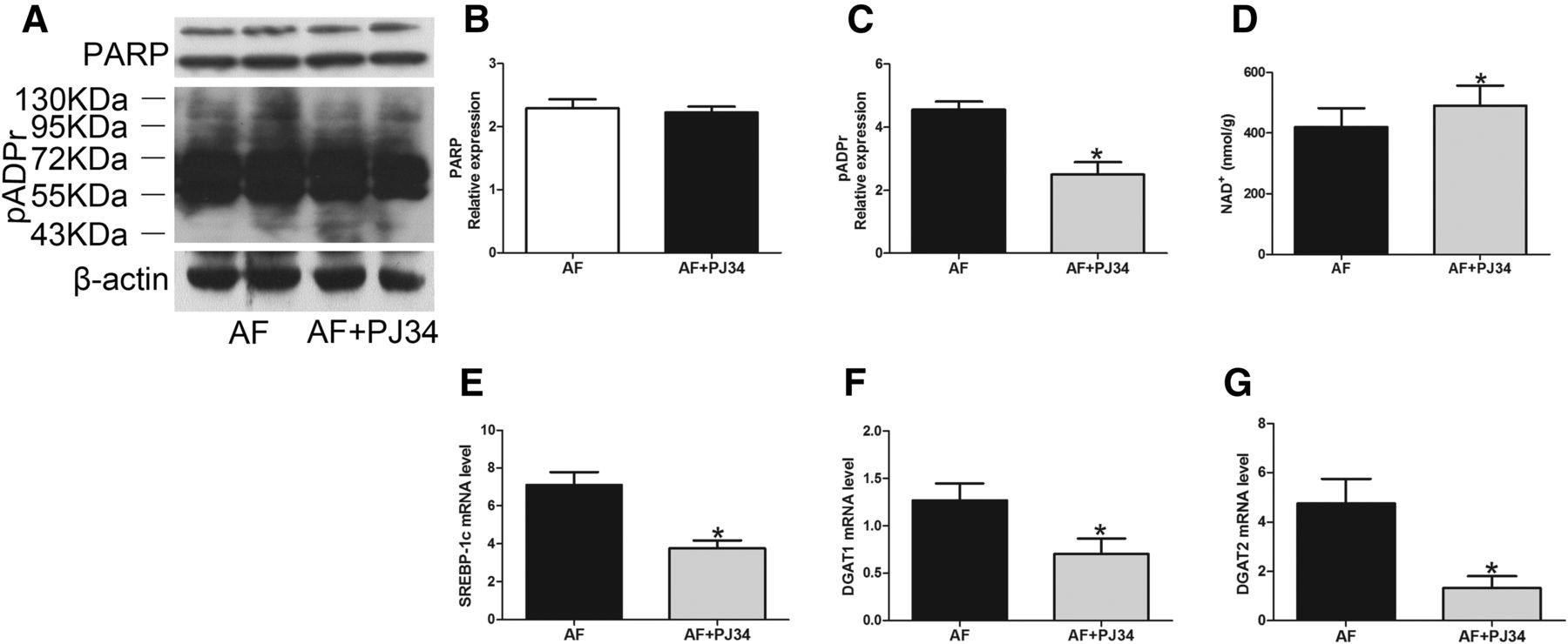
- Fig. 6.
PJ34 injection elevated hepatic NAD+ levels and lowered gene expression in TG anabolism in alcohol-fed mice. Male C57BL/6 mice were fed alcohol-containing liquid diets and injected intraperitoneally with or without PJ34 for 4 weeks. (A) Total proteins were extracted from liver tissues and were used to detect PARP expression and pADPr synthesis with specific antibodies. (B and C) PJ34 injection had no effect on PARP expression (B) and decreased pADPr synthesis (C). (D) PJ34 injection reduced hepatic NAD+ depletion compared with the AF group. (E–G) PJ34 injection significantly alleviated hepatic critical genes expression of SREBP-1 (E), DGAT1 (F), and DGAT2 (G) compared with the AF group. Values are means ± S.D. (n = 6 per group). *P < 0.05 (compared with the AF group).
- Fig. 7.
NR supplementation attenuated hepatic TG accumulation in alcohol-fed mice. Mice were fed alcohol-fed liquid diets with NR supplementation for 4 weeks. (A–C) NR supplementation augmented body weight (A) and decreased the liver/body weight rate compared with the AF group (B), whereas NR did not change the epididymal fat pad/body weight rate (C). (D and E) NR supplementation decreased hepatic TG content (D) and did not change hepatic TC content (E) compared with the AF group. (F) Supplementation with NR elevated hepatic NAD+ content in the AF + NR group. *P < 0.05 (significant differences compared with the AF group).


Additional Files
DATA SUPPLEMENT
- Supplemental Data -
Figure S1. Olaparib or Rucaparib injection attenuated hepatic fat droplet accumulation in alcohol-fed mice. C57BL/6 mice were fed with alcohol-containing liquid diet with/without Olaparib or Rucaparib intraperitoneal injection for four weeks. In comparison to the AF group, both Olaparib and Rucaparib injection significantly attenuated hepatic fat droplet accumulation in alcohol-fed mice. Liver sections were stained with H&E (x 400) (A). Relative quantitative statistics of fat droplet fold change (B). The data are expressed as the mean ± SD (n = 6 per group); # and & p < 0.05 compared to the AF group. * p < 0.05 compared to the PF group.PF: pair-fed; AF: alcohol-fed.
Figure S2. Olaparib and Rucaparib injection attenuated hepatic TG accumulation in alcohol feeding mice. After treated with/without Olaparib or Rucaparib intraperitoneal injection for four weeks. In comparison to the AF group, both Olaparib and Rucaparib injection augmented body weight (A). Rucaparib lowered liver/body weight rate (B). But Olaparib and Rucaparib injection had no significant change on epididymis fat/body weight rate (C). Olaparib and Rucaparib injection reduced the level of circulating ALT (D) and had no significant change on AST (E). Olaparib and Rucaparib intraperitoneal injection decreased hepatic TG content (F) and the level of circulating TG (G), but had no difference on hepatic TC content (H). Both of them significantly changed the level of circulating TC (I) in comparison to the AF group. The data are expressed as the mean ± SD (n = 6 per group); # and & p < 0.05 compared to the AF group. * p < 0.05 compared to the PF group. PF: pair-fed; AF: alcohol-fed; ALT: alanine transaminase; AST: aspartate transaminase;TC:cholesterol; TG: triglyceride.
Figure S3. Olaparib and Rucaparib injection elevated hepatic NAD+ level in alcohol-fed mice. Male C57BL/6 mice were fed with alcohol-containing liquid diets and injected intraperitoneally with/without Olaparib or Rucaparib for four weeks. Total proteins were extracted from liver tissues and used to detect the expression of PARP and the synthesis of pADPr (A) with specific antibodies. Olaparib and Rucaparib injection had no effect on PARP expression (B). Rucaparib decreased pADPr synthesis in alcohol-fed mice (C). Olaparib and Rucaparib injection reduced hepatic NAD+ depletion in comparison to the AF group (D). The data are expressed as the mean ± SD (n = 6 per group); # and & p < 0.05 compared to the AF group. * and @ p < 0.05 compared to the PF group. PF: pair-fed; AF:alcohol-fed; pADPr:polymeric adenosine diphosphate ribose; PARP: poly ADP ribose polymerase.
Figure S4. Increased PARP activity via treating with H2O2 reduced intracellular NAD+
level and enhanced intracellular TG content in hepatocyte. HepG2 cells were exposed to complete DMEM medium containing H2O2 (50 µM) for 24 h. The expression of intracellular PARP and pADPr were detected by Western blot (A). H2O2 treatment had no significant change on the expression of PARP (B) and elevated the synthesis of pADPr (C). H2O2 treatment decreased intracellular NAD+ content (D). Moreover, H2O2 treatment also increased intracellular TG content (E). Total RNAs were extracted from cells. The mRNA of critical genes was detected by RT-PCR. H2O2 treatment elevated intracellular SREBP-1c (F), DGAT1 (G), and DGAT2 (H) gene expression. All values were denoted as means ± SD from three or more independent batches of cells. * p < 0.05 compared to control group. pADPr: polymeric adenosine diphosphate ribose; PARP: poly ADP ribose polymerase; TG: triglyceride; DGAT1/2: diglyceride acyltransferase 1/2; SREBP-1c: sterol regulatory element binding protein 1c.
Figure S5. PJ34 injection decreased the levels of circulating pro-inflammatory cytokines in
alcohol-fed mice. PJ34 injection significantly decreased the levels of circulating MCP-1, IL-6 and TNF-α (A-C) in comparison to the alcohol-fed mice.
Figure S6. Pharmacological inhibition of PARP attenuated chronic alcohol-induced liver
inflammation in vivo. C57BL/6 mice were fed with alcohol-containing liquid diet with/without PARP inhibitor (Olaparib, Rucaparib or PJ34) intraperitoneal injection for four weeks. Immunohistochemical method was used to detect the expression of pro-inflammatory cytokines IL-6, MCP-1 and TNF-α in liver tissues (x 400) (A). In comparison to the PF group, Chronic alcohol consumption increased pro-inflammatory cytokines secretion. Olaparib, Rucaparib or PJ34 injection significantly attenuated pro-inflammatory cytokines secretion in alcohol-fed mice. Relative quantitative statistics of IL-6, MCP-1 and TNF-α fold change (B-D). The data are expressed as the mean ± SD (n = 6 per group); * p < 0.05 compared to the PF group. #, & and @ p < 0.05 compared to the AF group. PF: pair-fed; AF: alcohol-fed.
Figure S7. Chronic alcohol exposure induced hepatic SREBP-1 activation which attenuated by PJ34 injection. Total proteins were extracted from liver tissues and used to detect the expression of SREBP-1 (A and B) with specific antibody. SREBP-1 precursor (125KDa) and mature SREBP-1 (68KDa) were significantly increased in alcohol-fed mice (C). PJ34 injection reduced SREBP-1 expression in comparison to the AF group (D). The data are expressed as the mean ± SD (n = 6 per group); * p < 0.05 compared to the PF group. # p < 0.05 compared to the AF group. PF: pair-fed; AF:alcohol-fed; SREBP-1, sterol regulatory element binding protein 1.
- Supplemental Data -


