


Abstract
Spinal muscarinic acetylcholine receptors (mAChRs) play an important role in the regulation of nociception. To determine the role of individual mAChR subtypes in control of synaptic GABA release, spontaneous inhibitory postsynaptic currents (sIPSCs) and miniature IPSCs (mIPSCs) were recorded in lamina II neurons using whole-cell recordings in spinal cord slices of wild-type and mAChR subtype knockout (KO) mice. The mAChR agonist oxotremorine-M (3-10 μM) dose-dependently decreased the frequency of GABAergic sIPSCs and mIPSCs in wild-type mice. However, in the presence of the M2 and M4 subtype-preferring antagonist himbacine, oxotremorine-M caused a large increase in the sIPSC frequency. In M3 KO and M1/M3 double-KO mice, oxotremorine-M produced a consistent decrease in the frequency of sIPSCs, and this effect was abolished by himbacine. We were surprised to find that in M2/M4 double-KO mice, oxotremorine-M consistently increased the frequency of sIPSCs and mIPSCs in all neurons tested, and this effect was completely abolished by 4-diphenylacetoxy-N-methylpiperidine methiodide, an M3 subtype-preferring antagonist. In M2 or M4 single-KO mice, oxotremorine-M produced a variable effect on sIPSCs; it increased the frequency of sIPSCs in some cells but decreased the sIPSC frequency in other neurons. Taken together, these data strongly suggest that activation of the M3 subtype increases synaptic GABA release in the spinal dorsal horn of mice. In contrast, stimulation of presynaptic M2 and M4 subtypes predominantly attenuates GABAergic inputs to dorsal horn neurons in mice, an action that is opposite to the role of M2 and M4 subtypes in the spinal cord of rats.
Muscarinic acetylcholine receptor (mAChRs) in the dorsal horn of the spinal cord regulate different physiological functions, including nociception. Intrathecal administration of muscarinic agonists or acetylcholinesterase inhibitors produces a potent analgesic effect in many different species, including rats, mice, and humans (Iwamoto and Marion, 1993; Naguib and Yaksh, 1994; Hood et al., 1997; Ellis et al., 1999; Duttaroy et al., 2002; Chen and Pan, 2004). Molecular cloning studies have revealed five molecularly distinct mAChRs referred to as M1-M5 (Caulfield, 1993; Wess, 1996). The five mAChR subtypes are all linked to different types of G proteins. The odd-numbered muscarinic subtypes (M1, M3, and M5) are selectively linked to Gq/11 proteins, whereas the even-numbered subtypes (M2 and M4) are preferentially coupled to the pertussis toxin-sensitive Gi/o proteins (Felder, 1995; Wess, 1996; Caulfield and Birdsall, 1998). Receptor autoradiography and immunocytochemistry studies have shown that the highest density of muscarinic receptors in the spinal cord is located in the superficial laminae in both rats and humans (Yamamura et al., 1983; Scatton et al., 1984; Villiger and Faull, 1985; Hoglund and Baghdoyan, 1997; Li et al., 2002). Previous studies have documented that M2, M3, and M4 muscarinic receptor subtypes are present in the spinal cord dorsal horn (Hoglund and Baghdoyan, 1997; Yung and Lo, 1997; Duttaroy et al., 2002; Chen et al., 2005). Although the role of the M2 and M4 receptor subtypes in the muscarinic agonist-induced analgesia has been established (Ellis et al., 1999; Gomeza et al., 1999; Duttaroy et al., 2002; Li et al., 2002), the mechanisms by which each mAChR subtype contributes to the muscarinic analgesia in the spinal cord are not well understood.
The spinal lamina II neurons are tonically controlled by glutamatergic excitatory and GABAergic/glycinergic inhibitory inputs. Glutamate released from primary afferent nerves is a major excitatory neurotransmitter that conveys nociceptive information to the superficial lamina neurons (Yoshimura and Jessell, 1990). Our previous study in rats has shown that stimulation of mAChRs inhibits glutamate release and inhibition of glutamate release are indirectly mediated by increased GABA release and activation of presynaptic GABAB receptors (Li et al., 2002). In addition, muscarinic agonists probably produce analgesia through the inhibition of deep dorsal horn projection neurons of rats through GABAB receptors in rats (Chen and Pan, 2004). Thus, stimulation of spinal GABA release and GABAB receptors is considered an important mechanism by which muscarinic agonists exert their analgesic effects (Li et al., 2002; Chen and Pan, 2004).
Using a proper combination of mAChR subtype-preferring antagonists and the muscarinic toxin, we recently found that the M2, M3, and M4 receptor subtypes all contribute to the potentiation of GABAergic tone in the spinal cord of rats (Zhang et al., 2005). However, the currently available “selective” muscarinic antagonists are endowed with only a relatively small degree of subtype specificity (Wess, 1996; Caulfield and Birdsall, 1998). Therefore, it is essential to further substantiate the role of mAChR subtypes in the regulation of GABAergic inputs to spinal dorsal horn neurons using other approaches, such as mAChR subtype knockout (KO) mice (Wess, 2004). In the present study, we determined the role of individual mAChR subtypes in the regulation of synaptic GABA release to spinal lamina II neurons using mAChR KO mice. This study revealed an unexpected species difference in the specific role of individual mAChR subtypes in regulating GABAergic inputs to spinal dorsal horn neurons.
Materials and Methods
Animals. All wild-type and mAChR subtype single- and double-KO mice were obtained from University of Tokyo (from Minoru Matsui) and the National Institute of Diabetes and Digestive and Kidney Diseases (from Jürgen Wess). The generation and breeding of M2 KO, M4 KO, M3 KO, M1/M3 double-KO, and M2/M4 double-KO mice have been described elsewhere (Gomeza et al., 1999; Duttaroy et al., 2002; Matsui et al., 2002; Fukudome et al., 2004). All of the KO mice from Minoru Matsui originated from 129/SvJ ES cells and were backcrossed onto the C57BL/6JJcl background (CLEA Japan, Inc., Tokyo, Japan) for more than seven generations. Some of the M3 KO and M2/M4 double-KO mice were obtained from Jürgen Wess. The M3 KO mice of Jürgen Wess were backcrossed for 10 generations onto the C57BL/6NTac background. The M2/M4 double-KO mice of Jürgen Wess were maintained on a 129J1/CF1 mixed genetic background (Duttaroy et al., 2002). Age-(5-7 weeks old) and sex-matched descendants derived from the wild-type mice resulting from intercrossing of these mutants were used in this study. Because the effects of oxotremorine-M on sIPSCs and mIPSCs were identical in the two strains of wild-type, M3 KO, and M2/M4 double KO mice, the electrophysiology data from M3 KO and M2/M4 double KO mice were pooled. Mouse genotyping was carried out by Southern blotting and polymerase chain reaction analysis of mouse-tail DNA, as described previously (Duttaroy et al., 2002; Fukudome et al., 2004). The experimental protocols and procedures were approved by the Animal Care and Use Committee of the Pennsylvania State University College of Medicine and conformed to the guidelines of the National Institutes of Health Guide for the Care and Use of Laboratory Animals.
Spinal Cord Slice Preparations. Mice were anesthetized with 2% halothane in O2, and the lumbar segment of the spinal cord was rapidly removed through a limited laminectomy. Mice were then killed by inhalation of 5% halothane. The segment of the lumbar spinal cord was immediately placed in an ice-cold sucrose artificial cerebrospinal fluid (aCSF) presaturated with 95% O2 and 5% CO2. The sucrose aCSF contained 234 mM sucrose, 3.6 mM KCl, 1.2 mM MgCl2, 2.5 mM CaCl2, 1.2 mM NaH2PO4, 12.0 mM glucose, and 25.0 mM NaHCO3. Transverse spinal cord slices (350 μm) were cut in the ice-cold sucrose aCSF and then preincubated in Krebs solution oxygenated with 95% O2 and 5% CO2 at 34°C for at least 1 h before they were transferred to the recording chamber. The Krebs solution contained 117.0 mM NaCl, 3.6 mM KCl, 1.2 mM MgCl2, 2.5 mM CaCl2, 1.2 mM NaH2PO4, 11.0 mM glucose, and 25.0 mM NaHCO3.
The lamina II has a distinct translucent appearance and can be identified easily under the microscope. In this study, neurons in the outer zone of lamina II were selected for recording (Li et al., 2002; Pan et al., 2002). The neurons located in the dorsal one third of lamina II in the spinal slice were used for recording under a fixed-stage microscope (BX51WI; Olympus, Tokyo, Japan) with differential interference contrast/infrared illumination. The electrode for the whole-cell recordings was pulled from borosilicate glass capillaries using a puller (P-97; Sutter Instrument, Novato, CA). The impedance of the pipette was 4 to 7 MΩ when filled with internal solution containing 110 mM Cs2SO4, 5 mM KCl, 2.0 mM MgCl2, 0.5 mM CaCl2, 5.0 mM HEPES, 5.0 mM EGTA, 5.0 mM ATP-magnesium, 0.5 mM sodium-GTP, 1 mM guanosine 5′-O-(2-thiodiphosphate) (GDP-β-S), and 10 mM QX314, adjusted to pH 7.2 to 7.4 with 1 M CsOH (290-320 mOsm). GDP-β-S was added to the internal solution to block the possible postsynaptic effect mediated by muscarinic agonists through G proteins (Li et al., 2002; Zhang et al., 2005). QX314, a sodium-channel blocker, was added to the internal solution to suppress the action potential generation from the recorded cell. The slice was placed in a glass-bottomed chamber (Warner Instruments, Hamden, CT) and fixed with parallel nylon threads supported by a U-shaped stainless steel weight. The slice was continuously perfused with Krebs solution at 5.0 ml/min at 34°C maintained by an inline solution heater and a temperature controller (TC-324; Warner Instruments).
Electrophysiological Recordings. Recordings of postsynaptic currents were performed using the whole-cell voltage-clamp method, as described previously (Li et al., 2002; Zhang et al., 2005). Recordings of postsynaptic currents began 5 min later after whole-cell access was established and the current reached a steady state. The input resistance was monitored, and the recording was abandoned if it changed by more than 15%. Signals were recorded using an amplifier (MultiClamp 700A; Axon Instruments, Foster City, CA) at a holding potential of 0 mV, filtered at 1 to 2 kHz, digitized at 10 kHz, and stored in a Pentium computer with pCLAMP 9.0 (Axon Instruments). All spontaneous inhibitory postsynaptic currents (sIPSCs) were recorded in the presence of 2 μM strychnine, a glycine receptor antagonist, and 20 μM 6-cyano-7-nitroquinoxaline-2,3-dione, a specific glutamate non-N-methyl-d-aspartate antagonist. To record the miniature inhibitory postsynaptic currents (mIPSCs), 1 μM tetrodotoxin was added in the perfusion solution.
Oxotremorine-M, 4-diphenylacetoxy-N-methylpiperidine methiodide (4-DAMP), GDP-β-S, atropine, himbacine, strychnine, 6-cyano-7-nitroquinoxaline-2,3-dione, and bicuculline were obtained from Sigma (St. Louis, MO). Tetrodotoxin and QX314 were obtained from Alomone Labs (Jerusalem, Israel). Drugs were dissolved in Krebs solution and perfused into the tissue chamber using syringe pumps.
Data Analysis. Data are presented as means ± S.E.M. The sIPSCs and mIPSCs were analyzed offline with a peak detection program (MiniAnalysis; Synaptosoft, Decatur, GA). Measurements of the amplitude and frequency of sIPSCs and mIPSCs were performed over a period of at least 1 min during control, drug application, and recovery. For each analysis, at least 300 events were included. The sIPSCs and mIPSCs were detected by the fast rise time of the signal over an amplitude threshold above the background noise. Neurons were considered to be responsive to oxotremorine-M if the frequency or amplitude of sIPSCs and mIPSCs was altered >20%. The cumulative probability of the amplitude and interevent interval of sIPSCs and mIPSCs was compared using the Komogorov-Smirnov test, which estimates the probability that two cumulative distributions are similar. The effects of oxotremorine-M on the frequency and amplitude of sIPSCs and mIPSCs were determined by paired two-tailed Student's t test or repeated-measures analysis of variance. P < 0.05 was considered to be statistically significant.
Results
All lamina II neurons tested exhibited spontaneous IPSCs at a holding potential of 0 mV. There were no significant differences in the input resistance and the baseline frequency and amplitude of sIPSCs in different types of mAChR KO and wild-type mice (Table 1).
Comparison of the input resistance and the basal frequency and amplitude of sIPSCs in spinal lamina II neurons from the wild-type and different mAChR subtype KO mice
Effect of Oxotremorine-M on sIPSCs and mIPSCs in Wild-Type Mice. To determine the concentration-dependent effect of oxotremorine-M on sIPSCs in spinal cord lamina II neurons of wild-type mice, 1, 3, 5, and 10 μM oxotremorine-M, a nonselective agonist for all mAChR subtypes, was perfused to the tissue chamber. Each concentration was applied for a duration of 3 min. In a total of 15 lamina II neurons, 1 μM oxotremorine-M had no significant effect on the frequency and amplitude of sIPSCs. Oxotremorine-M from 3 to 10 μM significantly decreased the frequency of sIPSCs in 12 (80%) of 15 neurons in a concentration-dependent manner (Fig. 1, A-C). Oxotremorine-M decreased the amplitude of sIPSCs in 7 (58.3%) of the above 12 cells (from 15.76 ± 3.78 to 10.11 ± 1.37 pA at the concentration of 5 μM, P < 0.05) but did not significantly alter the IPSC amplitude in the remaining 5 cells (from 12.46 ± 1.12 to 11.44 ± 0.96 pA at the concentration of 5 μM). Oxotremorine-M had no significant effect on sIPSCs in the remaining three (20%) neurons. The sIPSCs were completely eliminated by 10 μM bicuculline, a specific GABAA receptor antagonist (Fig. 1A).
In another 17 lamina II neurons, the effect of 5 μM oxotremorine-M on mIPSCs was tested. Oxotremorine-M significantly decreased the frequency but not the amplitude of mIPSCs in 14 of 17 cells tested (Fig. 1D). Oxotremorine-M had no significant effect on the frequency (from 0.91 ± 0.18 to 0.88 ± 0.14 Hz) and amplitude (from 16.81 ± 1.65 to 15.80 ± 1.18 pA) of mIPSCs in the remaining three cells.
In 11 additional lamina II neurons, we tested the effect of the M2 and M4 subtype-preferring antagonist himbacine (Dorje et al., 1991; Miller et al., 1992; Doller et al., 1999) on oxotremorine-M-induced decreases in the frequency of sIPSCs in wild-type mice. In all 11 cells tested, 3 μM oxotremorine-M initially decreased the frequency of sIPSCs (Fig. 1E). Oxotremorine-M induced a large increase in the frequency of sIPSCs in the presence of 2 μM himbacine (Fig. 1E).

Effect of oxotremorine-M on sIPSCs and mIPSCs of spinal lamina II neurons in wild-type mice. A, original tracings of sIPSCs during control, application of 3 and 5 μM oxotremorine-M (Oxo), and washout in one lamina II cell. Note that the sIPSCs were abolished by 20 μM bicuculline. B, cumulative plot of sIPSCs of the same neuron in A showing the distribution of the interevent interval and amplitude during control, application of 5 μM oxotremorine-M, and washout. C, summary data showing that the effect of oxotremorine-M on the sIPSCs in 12 cells in wild-type mice. D, summary data showing the effect of oxotremorine-M on the frequency of mIPSCs in wild-type mice (n = 14). E, group data showing the effect of 3 μM oxotremorine-M on sIPSCs before and during the application of 2 μM himbacine (Him) in 11 lamina II neurons. Data are presented as means ± S.E.M. *, P < 0.05 compared with the control.
Effect of Oxotremorine-M on sIPSCs and mIPSCs in M1/M3 Double-KO and M3 Single-KO Mice. To assess the role of the M2 and M4 mAChR subtypes in the inhibitory effect of oxotremorine-M on synaptic GABA release, we tested the effect of oxotremorine-M on sIPSCs in M1/M3 double-KO and M3 single-KO mice. In M1/M3 double-KO mice, 3 to 10 μM oxotremorine-M significantly decreased the frequency of sIPSCs in a concentration-dependent manner in all 12 cells tested (Fig. 2A). Oxotremorine-M decreased the amplitude of sIPSCs in 6 of 12 cells (from 23.99 ± 4.45 to 16.32 ± 3.88 pA at the concentration of 5 μM, P < 0.05) but did not significantly alter the IPSC amplitude in the remaining 6 cells (from 16.68 ± 3.69 to 16.05 ± 4.19 pA at the concentration of 5 μM, P > 0.05).
In addition, we tested the effect of himbacine on oxotremorine-M-induced decreases in the frequency of sIPSCs in M1/M3 double-KO mice. In six cells tested, 3 μM oxotremorine-M initially decreased the frequency of sIPSCs (Fig. 2B). In the presence of 2 μM himbacine, oxotremorine-M had no significant effect on the frequency of sIPSCs (Fig. 2B).

Effect of oxotremorine-M on sIPSCs in M1/M3 double-KO and M3 single-KO mice. A, summary data showing the dose-response effect of oxotremorine-M on the frequency of sIPSCs in M1/M3 double-KO mice. B, group data showing the effect of 3 μM oxotremorine-M on sIPSCs before and during application of 2 μM himbacine (Him) in six lamina II neurons from M1/M3 double-KO mice. C, summary data showing the dose-response effect of oxotremorine-M on the frequency of sIPSCs in M3 single-KO mice. Data are presented as means ± S.E.M. *, P < 0.05 compared with respective controls.
Previous pharmacological studies have suggested the presence of the M1 subtype in the spinal dorsal horn (Iwamoto and Marion, 1993; Naguib and Yaksh, 1997). Thus, both M3 KO and M1/M3 double-KO mice were used to determine whether the M1 subtype plays a role in the effect of oxotremorine-M on synaptic GABA release in the spinal cord. In M3 single-KO mice, oxotremorine-M also decreased the frequency of sIPSCs in a concentration-dependent manner in all 15 cells tested (Fig. 2C). Oxotremorine-M decreased the amplitude of sIPSCs in 6 of 15 cells (from 22.68 ± 3.40 to 14.91 ± 2.52 pA at the concentration of 5 μM, P < 0.05) but had no effect on another 9 cells (from 10.98 ± 1.65 to 9.82 ± 1.58 pA at the concentration of 5 μM, P > 0.05). Application of 5 μM oxotremorine-M significantly decreased the frequency (from 1.15 ± 0.27 to 0.51 ± 0.11 Hz) but not the amplitude (11.82 ± 0.53 verses 11.28 ± 0.45 pA) of mIPSCs in 11 of 13 cells. Oxotremorine-M had no significant effect on the frequency (from 1.22 to 1.20 Hz in one cell and from 1.26 to 1.31 Hz in another cell) of mIPSCs in the remaining two cells in M3 single-KO mice.

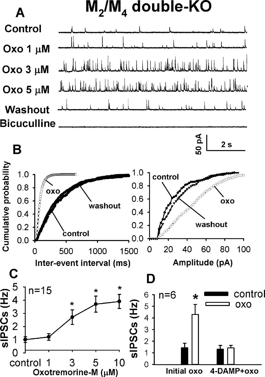
Effect of oxotremorine-M on sIPSCs in M2/M4 double-KO mice. A, raw tracings of sIPSCs during control, application of 1, 3, and 5 μM Oxo, and washout in one cell. Note that the sIPSCs were abolished by 20 μM bicuculline. B, cumulative probability plot of sIPSCs of the same neuron in A showing the distribution of the interevent interval and amplitude of sIPSCs during control, application of 5 μM oxotremorine-M, and washout. C, summary data showing the dose-response effect of oxotremorine-M on sIPSCs in 15 cells. D, effect of 3 μM oxotremorine-M on the frequency of sIPSCs in six cells before and after application of 25 nM 4-DAMP. Data are presented as means ± S.E.M. *, P < 0.05 compared with control.
Effect of Oxotremorine-M on sIPSCs and mIPSCs in M2/M4 Double-KO Mice. To determine the role of the M3 subtype in muscarinic regulation of synaptic GABA release to lamina II neurons, we then examined the effects of oxotremorine-M on the sIPSCs and mIPSCs in M2/M4 double-KO mice. It is noteworthy that oxotremorine-M (3-10 μM) significantly increased the frequency of sIPSCs in a concentration-dependent fashion in 15 (88%) of 17 neurons tested (Fig. 3, A-C). Oxotremorine-M had no effect on the frequency of sIPSCs in the remaining 2 of 17 neurons (from 1.06 Hz to 1.16 Hz and from 1.83 to 1.95 Hz at concentration of 5 μM). Oxotremorine-M also increased the amplitude of sIPSCs in 5 of 17 cells (from 17.62 ± 2.63 to 29.40 ± 6.71 pA at the concentration of 5 μM, P < 0.05) but had no significant effect on the amplitude of sIPSCs in the remaining 12 cells (from 23.18 ± 2.78 to 22.12 ± 2.65 pA at the concentration of 5 μM, P > 0.05). The sIPSCs were completely abolished by bath application of 10 μM bicuculline.
To determine whether the M3 subtype mediates the stimulating effect of oxotremorine-M on synaptic GABA release in M2/M4 double-KO mice, we further tested the effect of 3 μM oxotremorine-M on sIPSCs in the presence of 25 nM 4-DAMP, an M3 subtype-preferring muscarinic antagonist (Ehlert, 1996; Yigit et al., 2003; Zhang et al., 2005). The effect of 3 μM oxotremorine-M on sIPSCs was completely blocked by subsequent application of 25 nM 4-DAMP in all six cells examined (Fig. 3D).

Effect of Oxo on mIPSCs in M2/M4 double-KO mice. A, representative tracings of mIPSCs during control, application of 3 μM Oxo, and washout in one cell. B, cumulative probability plot of mIPSCs of the same neuron in A showing the distribution of the interevent interval and amplitude of mIPSCs during control, application of 3 μM oxotremorine-M, and washout. C, effect of 3 μM oxotremorine-M on the frequency of mIPSCs in 13 cells from M2/M4 double-KO mice. D, Effect of 3 μM oxotremorine-M on the frequency of mIPSCs before and after application of 25 nM 4-DAMP. These eight neurons were part of 13 cells shown in C. Data are presented as means ± S.E.M. *, P < 0.05 compared with the control.
In another 15 lamina II neurons from M2/M4 double-KO mice, 3 μM oxotremorine-M significantly increased the frequency but not the amplitude of mIPSCs in 13 neurons (P < 0.05, Fig. 4, A-C). This effect was also blocked by 25 nM 4-DAMP in all eight cells tested (Fig. 4D). Oxotremorine-M had no effect on the frequency (from 0.74 to 0.78 Hz and from 1.88 to 2.01 Hz) and amplitude (from 12.56 to 12.28 pA and from 27.16 to 30.94 pA) of mIPSCs in the remaining two cells.
Effect of Oxotremorine-M on sIPSCs in M2 Single-KO and M4 Single-KO Mice. To further delineate the role of the M2 and M4 receptor subtypes in the inhibitory action of oxotremorine-M on sIPSCs, the effect of oxotremorine-M on sIPSCs was tested in M2 single-KO and M4 single-KO mice, respectively. In M2 single-KO mice, 3 to 10 μM oxotremorine-M significantly increased the frequency of sIPSCs in a concentration-dependent manner in 16 (66.6%) of 24 neurons (Fig. 5, A-C). In contrast, oxotremorine-M had no significant effect on the frequency of sIPSCs in 4 (16.6%) of 24 neurons and significantly decreased the frequency of sIPSCs in 4 (16.6%) of 24 neurons. Oxotremorine-M increased the amplitude in 4 of the 24 cells (Fig. 5C) but had no significant effect on the amplitude of sIPSCs in 18 cells and even decreased the IPSC amplitude of 2 cells.
On the other hand, in M4 single-KO mice, 3 to 10 μM oxotremorine-M significantly increased the frequency of sIPSCs in only 4 (23.5%) of 17 neurons (Fig. 6). Oxotremorine-M had no significant effect on the frequency and amplitude of sIPSCs in 3 (17.7%, Fig. 6) of 17 neurons and significantly decreased the frequency and amplitude of sIPSCs in the remaining 10 (58.8%, Fig. 6B) of 17 neurons.

Effect of oxotremorine-M on the frequency of sIPSCs in M2 KO mice. A, original tracings of sIPSCs during control, application of 3 and 5 μM Oxo, and washout in one lamina II neuron. B, cumulative probability plot of sIPSCs of the same neuron in A showing the distributions of the interevent interval and amplitude during control, application of 5 μM oxotremorine-M, and washout. C, group data showing differential effects of 5 μM oxotremorine-M on the frequency of sIPSCs in 24 cells. Data are presented as means ± S.E.M. *, P < 0.05 compared with control.
Discussion
In this study, we used mAChR KO mice to define the role of individual mAChR subtypes in regulation of GABAergic inputs to spinal dorsal horn neurons. We found, unexpectedly, that oxotremorine-M decreased the frequency of GABAergic sIPSCs and mIPSCs in wild-type mice. Further studies showed that in M1/M3 double-KO and M3 single-KO mice, oxotremorine-M significantly decreased the frequency of sIPSCs and mIPSCs. It is noteworthy that oxotremorine-M significantly increased the frequency of sIPSCs and mIPSCs in M2/M4 double-KO mice. These results indicate that the M2 and M4 subtypes are responsible for the muscarinic inhibition of spinal GABA release in mice. On the other hand, stimulation of the M3 subtype is predicted to potentiate GABAergic tone in the spinal cord dorsal horn of mice. Therefore, this study provides new information that the activation of the M2 and M4 subtypes inhibits GABAergic inputs to spinal dorsal horn neurons in mice, which is opposite to the functional role of M2 and M4 mAChRs in the control of spinal GABA release in rats (Zhang et al., 2005). Nevertheless, stimulation of the M3 subtype potentiates synaptic GABA release in the spinal dorsal horn in both mice and rats.

Effect of oxotremorine-M on the frequency of sIPSCs in M4 KO mice. A, representative tracings of sIPSCs during control, application of 5 μM Oxo, and washout in one cell. B, group data showing differential effects of oxotremorine-M on the frequency of sIPSCs increased in 17 cells. Data are presented as means ± S.E.M. *, P < 0.05 compared with respective controls.
The mAChRs in the dorsal horn of the spinal cord are important for regulation of nociception. In this regard, intrathecal administration of muscarinic agonists or acetylcholinesterase inhibitors produces a potent analgesic effect in many different species, including rats, mice, and humans (Iwamoto and Marion, 1993; Naguib and Yaksh, 1994; Hood et al., 1997; Ellis et al., 1999; Duttaroy et al., 2002; Chen and Pan, 2004). Three mAChR subtypes, M2, M3, and M4 receptors, are present in the spinal cord dorsal horn, and the M2 and M3 subtypes are particularly concentrated in the superficial laminae of the spinal cord (Hoglund and Baghdoyan, 1997; Yung and Lo, 1997; Li et al., 2002). Because of a lack of highly specific agents for the individual mAChR subtypes, the receptor subtypes that mediate the analgesic effect of muscarinic agonists were not established until recently. The role of the M2 and M4 receptor subtypes in mediating the analgesic effect of mAChR agonists has been documented using M2 KO and M2/M4 double-KO mice (Gomeza et al., 1999; Duttaroy et al., 2002). The frequency of sIPSCs and mIPSCs is the most important measure of synaptic GABA release in this study. The change in the sIPSC amplitude by oxotremorine-M may reflect various pools of GABA-containing vesicles released from GABAergic terminals. We found in this study that oxotremorine-M inhibited sIPSCs and mIPSCs of dorsal horn neurons in wild-type mice, which is in contrast to the potentiating effect of oxotremorine-M on GABAergic IPSCs of dorsal horn neurons in rats (Zhang et al., 2005). In M3 single-KO and M1/M3 double-KO mice, oxotremorine-M consistently decreased the frequency of sIPSCs. Oxotremorine-M also significantly decreased the frequency, but not the amplitude, of mIPSCs in M3 KO mice, suggesting that the M2 and M4 subtypes are present on the presynaptic terminals of GABAergic interneurons in the mouse spinal cord. The role of M2 and M4 subtypes in the inhibition of synaptic GABA release in the mouse spinal dorsal horn is further supported by our finding that the M2 and M4 subtype-preferring antagonist himbacine reversed the inhibitory effect of oxotremorine-M on sIPSCs in wild-type mice and abolished the inhibitory effect of oxotremorine-M on sIPSCs in M1/M3 double-KO mice. Because oxotremorine-M consistently decreased the frequency of GABAergic sIPSCs and mIPSCs of dorsal horn neurons in wild-type mice, M2/M4 subtypes play a critical role in muscarinic inhibition of synaptic GABA release in the spinal dorsal horn of mice.
Contrary to the inhibitory effect of oxotremorine-M on IPSCs in wild-type and M3 KO mice, oxotremorine-M consistently increased the frequency of GABAergic sIPSCs in M2/M4 double-KO mice. Furthermore, the M3 subtype-preferring muscarinic antagonist 4-DAMP completely blocked the effect of oxotremorine-M on sIPSCs in M2/M4 double-KO mice. These data strongly suggest that activation of the M3 subtype potentiates synaptic GABA release in the spinal dorsal horn of mice. In wild-type mice, the role of the M3 subtype in muscarinic modulation of GABA release seems to be overwhelmed by the dominant action of the M2/M4 subtypes in the spinal dorsal horn of mice. This may explain why the potential role of the M3 subtype in muscarinic potentiation of spinal GABA release is only revealed by the use of M2/M4 double-KO mice and himbacine. Because oxotremorine-M significantly increased the frequency but not the amplitude of mIPSCs in M2/M4 double-KO mice, the M3 subtype is also probably located on the presynaptic terminals of GABAergic interneurons in the mouse spinal cord. The presence of the M3 receptor subtype in the rat spinal cord has been suggested in a radioligand binding study (Hoglund and Baghdoyan, 1997). We have shown that the M3 subtype plays a role in the potentiation of spinal GABA release by oxotremorine-M in rats (Zhang et al., 2005). However, the physiological function of the M3 subtype in the spinal cord remains largely unknown, and its functional role in the spinal analgesic effect of muscarinic receptor agonists needs to be further defined in M3 KO mice. Although previous pharmacological studies have suggested the presence of the M1 subtype in the spinal dorsal horn (Iwamoto and Marion, 1993; Naguib and Yaksh, 1997), we found no difference in the inhibitory effect of oxotremorine-M on sIPSCs between the M3 single-KO and M1/M3 double-KO mice. Hence, these data suggest that the M1 subtype is not involved in the muscarinic regulation of synaptic GABA release to spinal dorsal horn neurons.
The variable effect of oxotremorine-M on sIPSCs in the M2 and M4 single-KO mice most probably occurs because the recorded neurons receive different afferent inputs with distinct presynaptic mAChR subtypes. It is unlikely that this variability can be explained by the presence of different types of lamina II neurons because the effect of oxotremorine-M on sIPSCs is consistent in M1/M3 double-KO, M3 single-KO, and M2/M4 double-KO mice. This notion is further supported by our finding that after the blockade of the M2 and M4 subtypes with himbacine, oxotremorine-M consistently increased (but not decreased) the frequency of sIPSCs in wild-type mice. Because of the opposing functions of the M3 and M2/M4 subtypes in regulating spinal GABA release in mice (increase versus decrease in GABA release, respectively), the net effect of oxotremorine-M on synaptic GABA release depends on the distribution and the relative density of the M3 and M2/M4 subtypes on the nerve terminals that synapse with the recorded neuron. We observed that oxotremorine-M increased the frequency of sIPSCs in most (66.6%) lamina II neurons but decreased the IPSC frequency in some (16.6%) cells in M2 single-KO mice. These results suggest that there may be more M3 than M4 mAChRs on the presynaptic terminals of GABAergic neurons in the mouse spinal dorsal horn. On the other hand, oxotremorine-M can stimulate both M2 (to decrease GABA release) and M3 (to increase GABA release) subtypes in M4 single-KO mice. We found that oxotremorine-M decreased the frequency of sIPSCs in 58.8% cells, whereas it increased the IPSC frequency in 23.5% of the dorsal horn neurons tested in M4 single-KO mice. These data suggest that the M2 subtype plays a greater role than the M3 subtype in muscarinic regulation of spinal GABA release. Therefore, these mAChR subtypes contribute to a different extent (i.e., M2 > M3 > M4) to the muscarinic modulation of spinal GABA release in mice. These electrophysiological data are consistent with the receptor binding and behavioral studies showing that M2 is the predominant mAChR subtype in the mouse spinal cord (Gomeza et al., 1999; Duttaroy et al., 2002; Chen et al., 2005; Oki et al., 2005).

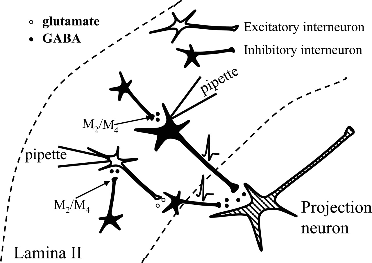
Diagram illustrating two possible mechanisms of inhibition of dorsal horn projection neurons as a result of decreased synaptic GABA release to lamina II interneurons by activation of M2 and M4 mAChR subtypes in the mouse spinal cord. See Discussion for details.
GABA is the most important inhibitory neurotransmitter in the central nervous system. Stimulation of spinal GABA release is an important mechanism by which muscarinic agonists produce their analgesic effects in the spinal dorsal horn of rats (Li et al., 2002; Chen and Pan, 2004). Nevertheless, we found that oxotremorine-M inhibits synaptic GABA release to dorsal horn neurons through the activation of M2 and M4 mAChRs in wild-type mice. Although it is uncertain how this effect (disinhibition) contributes to the mechanisms of spinal muscarinic analgesia in mice, it is possible that the location of mAChRs and spinal dorsal horn circuitry are organized differently in rats than in mice. As illustrated in Fig. 7, the mAChR agonists may inhibit dorsal horn projection neurons through at least two indirect mechanisms in mice. Assuming that the recorded lamina II neuron is an inhibitory interneuron (presumably GABAergic neurons), decreased GABA release after activation of the M2 and M4 subtypes could increase the excitability of the inhibitory interneuron. Therefore, it can potentiate the release of inhibitory neurotransmitters to dorsal horn projection neurons, resulting in the inhibition of nociceptive transmission. On the other hand, if the recorded lamina II neuron is an excitatory interneuron (supposedly glutamatergic, Fig. 7), decreased synaptic GABA release by mAChR agonists could increase the excitability of the excitatory interneuron. As a result, the increased synaptic glutamate release to the inhibitory interneuron that synapses directly with the dorsal horn projection neuron could lead to increased inhibitory tone to the dorsal horn projection neuron and suppression of nociceptive transmission in the mouse spinal cord. Our findings indicate that the pharmacological mechanisms through which mAChR subtypes regulate synaptic transmission and nociception at the spinal cord level are probably complex. For example, glutamate released from primary afferents to spinal dorsal horn neurons is important for transmission of nociceptive information (Yoshimura and Jessell, 1990; Li et al., 2002). The M2 and M4 subtypes may also be involved in the inhibition of glutamate release from primary afferent nerves to dorsal horn neurons (Li et al., 2002). The specific role of mAChR subtypes in regulating spinal glutamate release has not been examined in mice and will be determined in future studies.

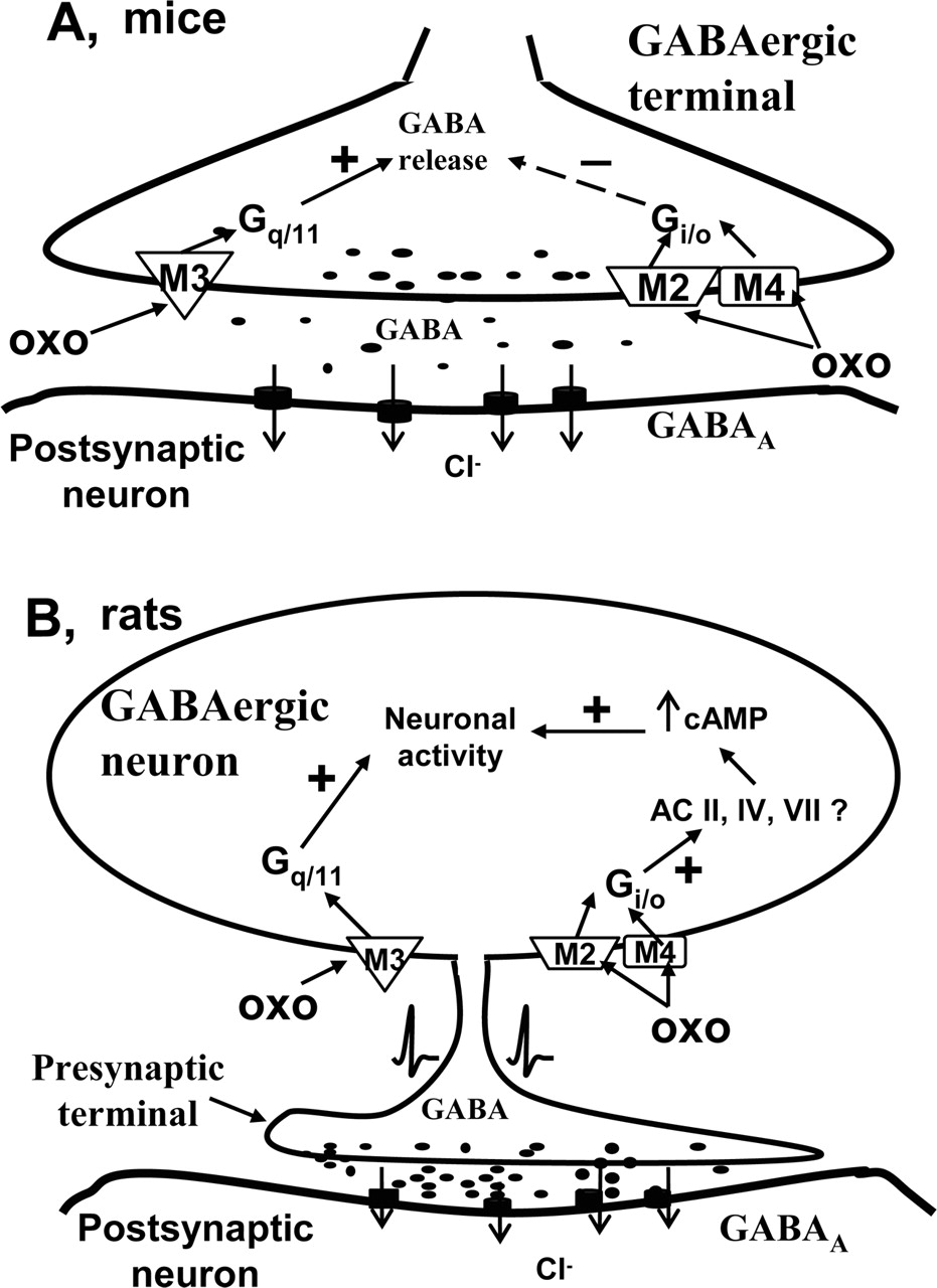
Schematic drawing highlighting the major differences in the subcellular location and physiological function of M2, M3, and M4 mAChR subtypes in control of synaptic GABA release to lamina II neurons in the spinal cord dorsal horn of mice (A) and rats (B). Note that M2, M3, and M4 mAChR subtypes may be located on the same or different GABAergic neurons and terminals. AC, adenylyl cyclase; -, inhibition; +, potentiation.
In summary, the most salient finding of the present study is that the activation of M2 and M4 mAChR subtypes in the mouse spinal cord inhibits GABA release (Fig. 8), which is opposite to the potentiating effect of the M2 and M4 subtypes on spinal GABAergic tone in rats (Zhang et al., 2005). Furthermore, unlike the rat spinal cord, in which the M2, M3, and M4 mAChRs are located on somatodendritic sites of GABAergic interneurons (Zhang et al., 2005), the mAChR subtypes (M2, M3, and M4) seem to be located on the presynaptic terminals of GABAergic interneurons in the spinal cord of mice (Fig. 8). The different subcellular location of the M2/M4 subtypes may be the basis for distinct functions and signaling mechanisms of these mAChR subtypes in regulating GABAergic synaptic transmission in the spinal dorsal horn of rats and mice. A species difference in the mAChR subtype signaling between rats and mice has been reported in the superior cervical ganglionic neurons (Shapiro et al., 1999, 2001). In this regard, the fast inhibition of N- and P/Q-type Ca2+ channels is mediated by the M4 subtype in the rat but by the M2 subtype in the mouse (Shapiro et al., 1999, 2001). The present study provides important new information about the species difference in regulation of GABAergic transmission by mAChR subtypes and the potential mechanisms of muscarinic analgesia in the spinal cord.
Footnotes
-
This study was supported by grants GM64830 and NS45602 from the National Institutes of Health (to H.-L.P.) and grant-in-aid for Scientific Research on Priority Areas 16067101 from the Ministry of Education, Culture, Sports, Science and Technology of Japan (to M.M.).
-
ABBREVIATIONS: mAChR, muscarinic acetylcholine receptor; sIPSC, spontaneous inhibitory postsynaptic current; mIPSC, miniature inhibitory postsynaptic current; GDP-β-S, guanosine 5′-O-(2-thiodiphosphate); 4-DAMP, 4-diphenylacetoxy-N-methylpiperidine methiodide; QX314, 2-((2,6-dimethylphenyl)amino)-N,N,N-triethyl-2-oxoethanaminium; aCSF, artificial cerebrospinal fluid; KO, knockout; Oxo, oxotremorine-M.
- Received August 14, 2005.
- Accepted December 19, 2005.
- The American Society for Pharmacology and Experimental Therapeutics


