Article Text

Abstract
BACKGROUND AND AIMS Stress may be an important factor in exacerbating inflammatory bowel disease but the underlying mechanism is unclear. Defective epithelial barrier function may allow uptake of luminal antigens that stimulate an immune/inflammatory response. Here, we examined the effect of chronic stress on colonic permeability and the participation of mast cells in this response.
METHODS Mast cell deficient Ws/Ws rats and +/+ littermate controls were submitted to water avoidance stress or sham stress (one hour/day) for five days. Colonic epithelial permeability to a model macromolecular antigen, horseradish peroxidase, was measured in Ussing chambers. Epithelial and mast cell morphology was studied by light and electron microscopy.
RESULTS Chronic stress significantly increased macromolecular flux and caused epithelial mitochondrial swelling in +/+ rats, but not in Ws/Ws rats, compared with non-stressed controls. Stress increased the number of mucosal mast cells and the proportion of cells showing signs of activation in +/+ rats. No mast cells or ultrastructural abnormalities of the epithelium were present in Ws/Ws rats. Increased permeability in +/+ rats persisted for 72 hours after stress cessation.
CONCLUSIONS Chronic stress causes an epithelial barrier defect and epithelial mitochondrial damage, in parallel with mucosal mast cell hyperplasia and activation. The study provides further support for an important role for mast cells in stress induced colonic mucosal pathophysiology.
- epithelial transport
- mast cells
- mitochondria
- psychological stress
- ultrastructure
Abbreviations used in this paper
- G
- conductance
- HRP
- horseradish peroxidase
- IBD
- inflammatory bowel disease
- Isc
- short circuit current
- Ws/Ws
- mast cell deficient rats with gene deletion at the “white spotting” (Ws) locus
- +/+
- normal littermates of Ws/Ws rats
Statistics from Altmetric.com
Although a controversial issue, clinical observations indicate that stress may be an important factor in exacerbating inflammatory bowel disease (IBD).1 One mechanism by which stress may influence intestinal disorders is by altering epithelial and immune responses in the gut.2 Indeed, experimental evidence has demonstrated reactivation or worsening of colonic inflammation after repeated exposure to stress.3-5 Epithelial cells participate in the regulation of mucosal inflammation by acting as a physical and functional barrier that limits the uptake of luminal antigens and pathogens. Among immune cells, mast cells play an important role in the regulation of epithelial transport in both human6 ,7 and rodent intestine,8 ,9 and it is widely accepted that nerve-mast cell interactions are involved in intestinal epithelial dysfunction.10 ,11 We and others have shown that nerves and mast cells participate in the development of colonic epithelial abnormalities induced by acute stress.12 ,13 The mast cell stabiliser doxantrazole abolished acute stress induced ion secretion and permeability enhancement in rat colon,12 and in a study using mast cell deficient mice, colonic mucin release in response to restraint stress was found to be mast cell dependent.13
Experiments involving repeated exposure of rodents to mild stress are commonly used as models of depression (reviewed by Willner14). These more chronic models better reflect the pattern of daily stress experienced by humans. In a recent study,15 we developed a rat model of ongoing psychological stress involving repetitive application of a mild stressor, water avoidance stress, for five consecutive days. The stressed rats reduced their food intake and lost weight during the five day period. In addition, jejunal conductance and macromolecular permeability were increased in stressed wild-type rats but not in mast cell deficient rats, suggesting an important role for mast cells in stress induced small bowel mucosal pathophysiology.
A recent clinical study showed that long term stress is more important than acute stressful events for the risk of exacerbation of ulcerative colitis.16 A possible mechanism could be stress induced deterioration in colonic barrier function. However, the effect of chronic stress on colonic epithelial physiology has not been reported. The specific aims of the present study were: (1) to investigate the effects of chronic stress on epithelial ion secretion and barrier function in rat colon; (2) to determine if mast cell activation plays a role in stress induced colonic mucosal responses; and (3) to investigate if colonic physiological abnormalities were accompanied by ultrastructural changes in colonocytes.
Methods
ANIMALS
Ws/Ws rats and their +/+ littermate controls were obtained from our breeding colony at McMaster University (original breeders obtained from Dr Y Kitamura, Osaka, Japan). Ws/Ws rats have a 12 base deletion in the tyrosine kinase domain of the c-kitgene and by 10 weeks of age no mast cells can be detected in their intestine, whereas +/+ rats have normal numbers of mast cells.17 Early in life, Ws/Ws rats also have anaemia, but this is largely corrected in rats older than 10 weeks of age.17 Weight (250–300 g) and age (14–20 weeks) matched rats, individually housed, were maintained on a 12:12 hour (8 am/8 pm) dark/light cycle and provided with food and water ad libitum. The Animal Care Committee at McMaster University approved all procedures.
STRESS PROTOCOL
Four groups of rats were studied: Ws/Ws rats, either stressed or sham stressed, and +/+ rats, either stressed or sham stressed. The rats were handled daily for two weeks by the same examiner and then submitted to water avoidance stress or sham stress daily for one hour over five days. The procedure involved placing the animals on a platform (8×6 cm) in a plastic container (56×50 cm) filled with warm water (25°C) to 1 cm below the platform. Sham stressed rats were placed on the same platform above a waterless container. The procedures were performed between 8:00 am and 10:00 am to minimise the effect of circadian rhythm. In previous experiments, we determined that the effects of acute stress on epithelial function were maximal at 5–7 hours after stress and returned to normal at 24 hours (Saunders and Perdue, unpublished observations). Based on this finding, six hours after the last stress or sham session, rats were killed by decapitation and tissues were obtained for functional and morphological studies.
Additional experiments were performed to assess the duration of stress effects on epithelial physiology. Since only +/+ rats showed stress induced epithelial abnormalities, an additional group of +/+ rats was submitted to the sham or stress protocols (five days) and then returned to their cages where they remained undisturbed. Rats were killed on days 1, 3, and 7 (n=8 for each time point) after completion of sham/stress protocols, and colonic tissue obtained for studies.
USSING CHAMBER STUDIES
Distal colonic segments were immediately immersed in oxygenated Krebs solution, stripped of longitudinal muscle and myenteric plexus, and 2–4 adjacent pieces from each segment were mounted in Ussing chambers (World Precision Instruments, Narco Scientific, Mississauga, Ontario, Canada). The chamber exposed 0.6 cm2 of tissue surface area to 8 ml of circulating oxygenated Krebs buffer at 37°C. The buffer contained (in mM) 115 NaCl, 1.25 CaCl2, 1.2 MgCl2, 2.0 KH2PO4, and 25 NaHCO3 (pH 7.35). The serosal buffer also contained 10 mM glucose as an energy source, osmotically balanced by 10 mM mannitol in the mucosal buffer. The chambers contained agar-salt bridges to monitor the potential difference across the tissue and to inject the required short circuit current (Isc) to maintain a zero potential difference as registered via an automated voltage clamp (Narco Scientific). Every five minutes, a pulse of 1 mV was passed through the tissue, and by measuring the change in Isc, the conductance was calculated by Ohm's law. Baseline values for Isc and conductance were recorded at equilibrium, 20 minutes after mounting of the tissues. Segments with signs of poor viability—that is, unstable Isc and/or conductance—were excluded from the study.
Mucosal to serosal transport of macromolecules was assessed by measuring transepithelial flux of horseradish peroxidase (HRP), a model protein antigen.18 Fifteen minutes after mounting the tissues, HRP (type VI, Sigma Chemical Co., St Louis, Missouri, USA) was added to the luminal buffer at a final concentration of 10−5 M and allowed to equilibrate for 30 minutes. Serosal samples (0.5 ml) were obtained at 30 minute intervals for two hours and replaced by 0.5 ml of appropriate buffer solution. HRP activity was determined by a modified Worthington method, as previously described.18 The mucosal to serosal flux of HRP was reported as the average value of two consecutive stable flux periods (between 30 and 90 minutes), and expressed as pmol/h/cm2.
HISTOLOGY
Full thickness segments of colon were fixed in Carnoy's fixative and stained with toluidine blue (Sigma). Overall morphology was assessed. Mast cells were counted in coded sections at 400× magnification using a micrometer grid (0.032 mm2 area). For each rat, 12 contiguous non-overlapping mucosal areas above themuscularis mucosae were evaluated.
ELECTRON MICROSCOPY
Colonic tissues were obtained from rats immediately after being killed by decapitation and also from chambers two hours after addition of HRP into the luminal compartment. Tissues were immediately fixed in 2.5% glutaraldehyde in 0.1 M sodium cacodylate buffer (pH 7.4) for two hours at 22°C, rinsed for 18 hours (4°C) with 0.05 Tris buffer (pH 7.6), and washed three times, five minutes each time. Methods for HRP product identification have been described previously.18Quantitative analysis of HRP uptake in intracellular endosomes and paracellular HRP transport were performed on coded high magnification photomicrographs, 15 per rat (four rats/group). The total area of HRP containing endosomes within colonocytes was determined in an area of 300 μm2 in the apical region of the cells, using a computerised image analysis system (Kontron Mop Videoplan; Kontron, Eching, Germany). Tissues were also examined for evidence of paracellular HRP transport in coded photomicrographs that were randomly selected. In each group, 1000 paracellular regions from four different rats were examined. HRP was identified as electron dense material within the paracellular regions (between lateral cell membranes of two adjacent cells).
Ultrastructural epithelial damage was evaluated in colonocytes by the presence of mitochondrial changes such as enlargement, swelling, disorientation, and disappearance of cristae. The mean area of individual mitochondria within the apical region of the colonocytes was measured in four rats per group, 15 sections per rat, using a computerised image analysis system as described above.
Mast cells were evaluated for signs of activation by electron microscopy and image analysis. Patterns of mast cell activation/degranulation were assessed as: (a) piecemeal degranulation, defined as loss of granule density without granule-cell membrane fusion,19 and (b) anaphylactic degranulation, defined as solubilisation of granular contents with fusion of intergranular and granule-cell membranes.20 Fifteen sections containing mast cells from each tissue (four rats per group) were randomly selected, the number of intracytoplasmatic granules counted, and each granule analysed for loss of density and perigranular vacuolisation.
FAECAL OUTPUT
At the end of each experimental session (stress or sham stress), the number of faecal pellets expelled was counted and recorded. Changes in faecal pellet output has previously been shown to be an index of changes in colonic transit time during stress in rats.21
STATISTICS
Results are expressed as mean (SEM). Results were analysed by two way ANOVA with contrasts to show the simple effects of the following main factors: treatment group (stress vsham) and strain (Ws/Ws v +/+). A between-within multivariate ANOVA, where the third factor was a repeated measures factor (time), was used to compare means at different days in strain and treatment groups. The unpaired Student'st test was used to compare post-treatment differences (at each time) on epithelial parameters in sham and stress groups, with Bonferroni correction for multiple testing. The Mann-Whitney U test and Fisher's exact test were also used when appropriate. p<0.05 was considered significant.
Results
EFFECTS OF STRESS ON ELECTROPHYSIOLOGY
After sham stress, baseline Isc was similar in colonic tissues from +/+ and Ws/Ws rats. Stress doubled the Isc in +/+ rats but had no effect in Ws/Ws rats compared with the respective non-stressed controls (fig 1A). The increased Isc in +/+ rats was significant, as shown by the treatment by strain interaction (F1,20=8.7; p<0.01).
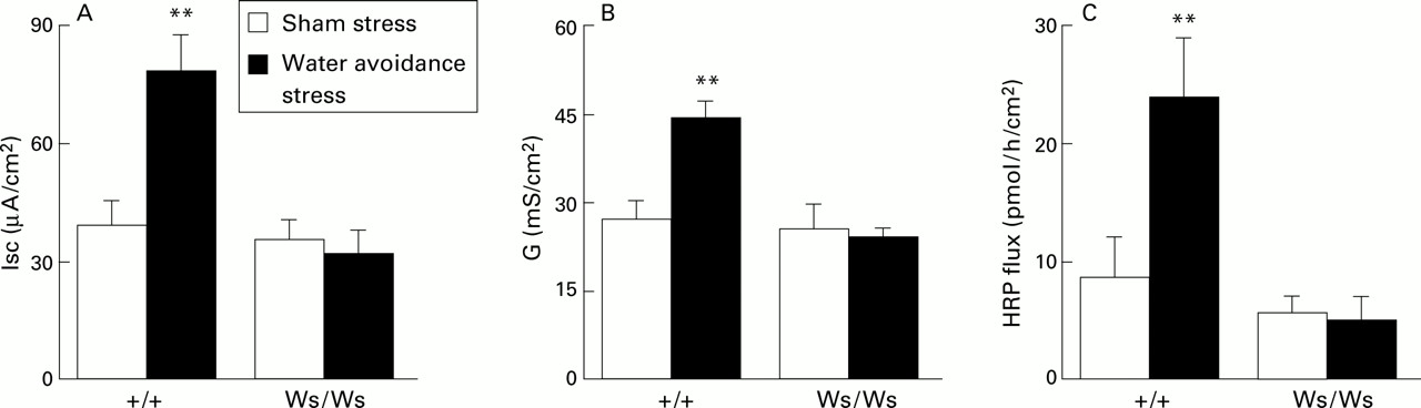
Effect of chronic stress on colonic physiology and macromolecule permeability. Mast cell deficient rats (Ws/Ws) and their normal mast cell containing littermates (+/+) were submitted to sham stress or water avoidance stress for five consecutive days. (A) Baseline short circuit current (Isc), (B) conductance (G), and (C) horseradish peroxidase (HRP) flux studied in Ussing chambers six hours after the final sham/stress session. Bars represent mean (SEM); n=6 rats/group, with 2–4 tissues averaged per rat. **p<0.01 v all other groups.
Similarly, conductance was not different in sham stressed +/+ and Ws/Ws rats whereas stress markedly increased colonic conductance in +/+ rats (F1,20=19.3; p<0.001) but not in Ws/Ws rats (fig 1B)
EFFECTS OF STRESS ON MACROMOLECULAR TRANSPORT
HRP flux across colonic tissues was similar after sham stress in +/+ (8.9 (3.3) pmol/h/cm2) and Ws/Ws (5.9 (1.4) pmol/h/cm2) rats. Stress enhanced HRP flux across the colonic epithelium in +/+ rats threefold to 24.3 (4.9) pmol/h/cm2 (F1,20=8.1; p<0.01) (fig 1C) whereas no increase was found in Ws/Ws rats.
On electron microscopy, HRP was evident within endosomes in colonocytes of both stressed and control rats. However, the number and size of HRP containing endosomes were significantly higher in stressed +/+ rats compared with the three other groups, as indicated by the HRP-endosome area (table 1, fig 2A, B). In addition, HRP was observed within colonic tight junctions and paracellular regions only in stressed +/+ rats, where 30% of paracellular spaces contained HRP (fig2B).
Electron microscopy findings in the colonocytes of mast cell deficient rats (Ws/Ws) and normal littermates (+/+) after chronic stress or sham stress.
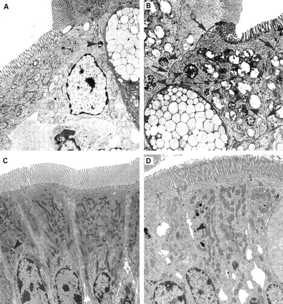
Effect of chronic stress on horseradish peroxidase (HRP) transport and epithelial mitochondria. Mast cell deficient rats (Ws/Ws; left) and their normal mast cell containing littermates (+/+; right) were submitted to water avoidance stress (top photomicrographs) or to sham stress (bottom photomicrographs) for five consecutive days. (A) Photomicrograph showing an HRP filled endosome (large arrowhead) in a colonocyte of a stressed Ws/Ws rat. Paracellular passage of HRP and mitochondrial abnormalities were not observed in Ws/Ws rats. (B) Larger and more numerous HRP filled endosomes (arrowheads) as well as HRP within tight junctions and paracellular spaces between colonocytes (arrows) were observed in the colon of stressed +/+ rats, as shown in this photomicrograph. Epithelial damage is demonstrated by swollen mitochondria with loss of cristae. (C, D) Colonocytes with small endosomal uptake of HRP (arrowheads) and normal mitochondria from sham stressed Ws/Ws (C) and +/+ rats (D), respectively. Mitochondrial surface area was significantly increased in stressed +/+ rats compared with the three other groups (table 1). Magnification: ×3000. Each photomicrograph is representative of four rats/group and 15 sections/colonic tissue.
EFFECTS OF STRESS ON EPITHELIAL MORPHOLOGY
On light microscopy, no colonic mucosal abnormalities were observed in either rat group after exposure to stress. However, using electron microscopy, ultrastructural signs of epithelial damage were observed in colonic tissues from all stressed +/+ rats. Colonocytes in these rats showed enlarged mitochondria with loss of cristae (fig 2C, D) whereas this abnormality was not found in cells of sham stressed +/+ or Ws/Ws rats or in cells of stressed Ws/Ws rats (table 1).
EFFECTS OF STRESS ON MUCOSAL MAST CELLS
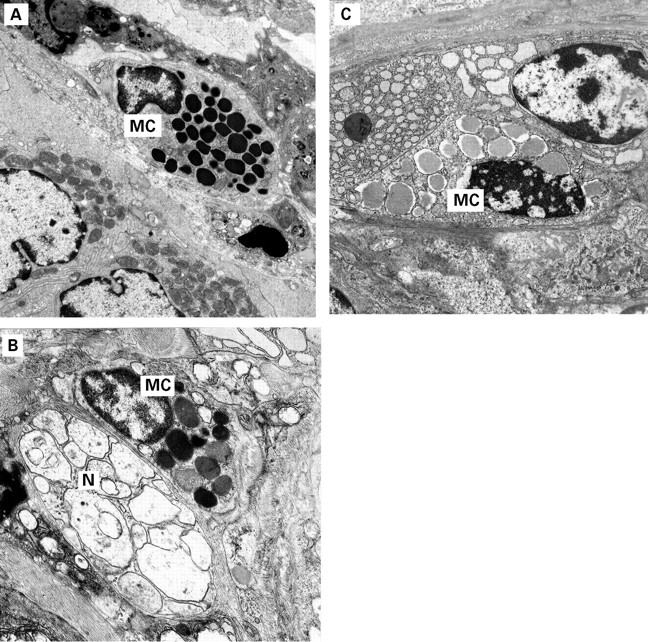
Mast cells were completely absent in the colon of Ws/Ws rats. In +/+ rats, stress exposure for five days was associated with the appearance of a significantly higher number of mast cells in the mucosa of the colon compared with the sham stressed group (table 2). In addition, as assessed by electron microscopy, a higher proportion of mucosal mast cells (∼30%) in tissues from stressed +/+ rats had granules with signs of activation (table 2) whereas changes in granule morphology were observed only in a minority of mast cells (6%) in the mucosa of sham stressed rats. Piecemeal-type degranulation, indicated by loss of granular density, was the predominant pattern observed in activated mast cells, while ultrastructural evidence of anaphylactic-type degranulation or mixed patterns were occasionally observed (fig 3A–C). Frequently, mast cells showing piecemeal-type degranulation were in close proximity to nerves (fig3B).
Effects of stress on mast cell numbers and activation

Effect of chronic stress on mast cells in +/+ rats. (A) A non-activated mast cell (MC) in the mucosa of a sham stressed +/+ rat showing numerous homogeneous electron dense granules. (B) A mast cell (MC) with piecemeal-type degranulation. The mast cell is in close apposition to an emptied nerve terminal (N) in the mucosa of a stressed +/+ rat, and shows loss of intragranular density without fusion of intergranular membranes, retained oval granules in the cytoplasm, and absence of extruded granules contents or granule membranes, indicative of piecemeal-type degranulation. (C) A mast cell (MC) in the mucosa of a stressed +/+ rat showing enlarged granules with solubilisation of intragranular contents, fusion of intergranular membranes (arrows), and absence of extruded material, indicative of a mixed anaphylactic-piecemeal degranulation pattern. Magnification: A–C, ×3000.
EFFECTS OF STRESS ON FAECAL OUTPUT
Defecation was increased during exposure to stress, as shown by the higher number of faecal pellets expelled by stressed versus sham stressed rats. This effect was significant for both +/+ (10.6 (0.9) pellets/h) and Ws/Ws (12.4 (0.9)) stressed rats compared with their controls (+/+: 1.0 (0.1) pellets/h, F1,36=95; Ws/Ws: 1.0 (0.2), F1,36=146; p<0.001 for both).
DURATION OF STRESS EFFECTS ON EPITHELIAL FUNCTION
Stress induced enhancement of HRP flux in +/+ rats continued to be significantly (p<0.05) elevated on days 1 (16.2 (1.1) pmol/h/cm2, n=4) and 3 (10.6 (1.2) pmol/h/cm2, n=4) after cessation of stress compared with tissues from sham treated rats on days 1 (6.4 (1.1) pmol/h/cm2, n=4) and 3 (6.6 (0.5) pmol/h/cm2, n=4), respectively. However, by day 7 after stress, HRP fluxes in the colon were similar in both groups (7.1 (0.5) and 6.4 (0.8) pmol/h/cm2 in stressed and sham rats, respectively, n=4 in each group).
Discussion
We have shown that repetitive exposure to water avoidance stress, a psychological stressor, induced sustained abnormalities in colonic epithelial barrier function, and that mast cells played an important role in this response. Participation of mast cells in intestinal epithelial dysfunction has been documented in several rodent models, such as immediate hypersensitivity,8 ,9 acute stress,12 Clostridium difficileinfection,22 as well as in inflamed human colon.6 We have previously shown that in the rat colon, a single exposure to acute stress stimulates epithelial ion secretion and increases transepithelial transport of macromolecules.12However, a model of more chronic stress, rather than acute stress, may better represent ongoing exposure to stressful life events in humans. Information regarding the effects of chronic stress on gut epithelial function and the role of mast cells is scarce, particularly in the colon. A recent study documented that the epithelial secretory response to 5-hydroxytryptamine in the distal colon was altered after three days of immobilisation for two hours/day,23 and chronic stress was also shown to stimulate colonic epithelial cell proliferation.24 In the present study, we report for the first time that chronic stress markedly increased HRP flux indicating enhanced epithelial permeability to macromolecules in the rat distal colon. Moreover, enhanced HRP flux involved both transcellular and paracellular routes across the epithelium. Baseline Isc, indicating net ion secretion, was also increased by chronic stress. A key finding was that these epithelial abnormalities were manifested only in +/+ rats with normal mucosal mast cell numbers, suggesting that mast cells are required for expression of stress induced epithelial transport abnormalities in the rat colon.
In parallel with abnormalities of epithelial function, we found ultrastructural evidence of mitochondrial changes in colonocytes of stressed +/+ rats whereas Ws/Ws mast cell deficient rats did not develop any changes in epithelial morphology. In the gastrointestinal tract, to our knowledge, mitochondrial abnormalities have only been reported in enterocytes of rats after 24 hours of restraint stress.25 It is known that certain severe stressors such as activity stress can induce overt damage involving bleeding and thinning of the colonic mucosa.26 However, our finding that a mild chronic stressor provoked ultrastructural changes of the epithelium, but only in tissues with mast cells, is a novel observation. Similar mitochondrial changes in intestinal epithelia have been induced by uncoupling of oxidative phosphorylation and associated with an increased epithelial permeability,27 ,28 involving rearrangement of F-actin and tight junction proteins.29Swelling of mitochondria may lead to rupture of the outer mitochondrial membrane and release of cytochrome c, a strong activator of the caspases which trigger the cell to undergo apoptosis.30However, depending on the cellular concentration of ATP and oxygen radicals, cell necrosis could also follow. Epithelial events following mitochondrial damage found in our model of chronic stress need further investigation, and such studies are underway in our laboratory.
Ws/Ws rats have a 12 base deletion in the tyrosine kinase domain of the c-kit gene that results in lack or deficiency of melanocytes, erythrocytes, interstitial cells of Cajal in the myenteric plexus, and mast cells.17 ,31 Mast cells have been reconstituted in mast cell deficient mice but due to the heterogeneous genetic background of the Ws/Ws and +/+ rats (F2 generation of two inbred rat strains), reconstitution is not feasible in Ws rats because of cell rejection. Therefore, we cannot rule out other phenotypic abnormalities as contributors to the observed responses in Ws/Ws rats. However, the fact that Ws/Ws rats show normal intestinal epithelial responses to carbachol and forskolin in the presence of neural blockade (Berin and Perdue, unpublished results), together with pharmacological evidence of mast cell involvement in stress responses in the colon,12 ,13highlight the role of mast cells in the colonic response to stress and make unlikely a large contribution of other phenotypic abnormalities.
Our study showed that stress induced mast cell hyperplasia in the colonic mucosa of +/+ rats. An increase in mast cell numbers has previously been shown in the rodent brain after chronic psychological stress.32 The presence of increased numbers of mast cells may indicate either local hyperplasia of resident mast cells or influx of newly immigrated mast cells from bone marrow or the submucosa or, most probably, a combination of both processes. Although our study did not explore the mechanism accounting for mast cell hyperplasia, several molecules such as interleukin 3, interleukin 4, nerve growth factor, or stem cell factor promote mast cell proliferation. Of these, nerve growth factor has been shown to be released during stress,33 making it a possible candidate for mast cell hyperplasia in our system. In addition, activated mast cells are known to release a host of cytokines/chemokines and in fact to stimulate recruitment of additional mast cells into the tissue.34Whether mast cell hyperplasia represents a direct response to stress mediators or a secondary phenomenon to stress induced epithelial damage remains to be elucidated.
We and others have shown that acute stress results in mast cell activation, assessed by either release of specific mediators or by histological criteria in the human jejunum7 and rat brain.35 Here, we demonstrated that chronic stress increased the number of mucosal mast cells fivefold, as well as the degree of activation, as indicated by ultrastructural changes. In some mast cells we observed a mixed pattern of piecemeal-type and anaphylactic-type degranulation. However, the majority of activated mast cells showed the pattern of piecemeal-type degranulation, as indicated by loss of granular density without fusion of intergranular membranes and no signs of compound exocytosis.19 This latter pattern has been reported in the brain of rats submitted to stress35 as well as in the gastrointestinal tract of IBD patients.36 Although the significance of these morphological changes is unclear, they have been noted primarily, as in our study, in mast cells which are in close proximity to nerve fibres,20 ,35 and may represent either an event preceding exocytosis or nerve mediated activation.37 Release of histamine without apparent signs of exocytosis in rat intestinal mast cells has previously been described after electrical stimulation of the vagus nerve.38
Increased colonic motor activity is a well known effect of different types of acute stress.39 In acute restraint stress, changes in colonic smooth muscle motility to facilitate colonic transit and evacuation have been found to be correlated with faecal pellet output.21 In contrast with changes in epithelial function, we found no difference between +/+ and Ws/Ws rats in the stress induced increase in faecal pellet expulsion. This is in agreement with a previous study of immobilisation stress in mast cell deficient mice,13 and suggests that motor disturbances induced by stress involve pathways less dependent on mast cells than those inducing epithelial dysfunction.
Finally, as in the jejunum,15 enhanced colonic permeability to macromolecules lasted for three days after stress exposure. Persistent abnormalities in barrier function could have implications for the development of intestinal inflammation. Patients with Crohn's disease have increased intestinal permeability.40 This barrier dysfunction appears to precede mucosal inflammation,41 and recurrence of inflammation can be avoided by diversion of the faecal stream.42 In gene engineered models of IBD, germ free rodents do not develop intestinal inflammation.43Increased uptake of luminal microbial antigens may be required to activate mucosal immune cells leading to an inflammatory response involved in the pathogenesis and/or reactivation of IBD. Whether increased colonic permeability and epithelial damage induced by stress, as in this study, eventually leads to intestinal inflammation remains to be elucidated.
In summary, in the present study we have shown that chronic stress caused prominent and lasting alterations in epithelial function in the rat colon and that the stress induced enhanced epithelial antigen uptake was mast cell mediated. The study provides further support for the role of mast cells, not only as key cells in the regulation of gut epithelial physiology, but also as effectors in stress induced colonic permeability. Stress has been linked to the course of colonic inflammation,1-5 and mast cells, which play a key role in colonic epithelial responses to stress, are also implicated in the pathogenesis of intestinal inflammation.22 Therefore, mast cells may represent a therapeutic target in the management of stress related mucosal pathophysiology in the gastrointestinal tract.
Acknowledgments
Supported by grants from the Medical Research Council of Canada, the Academia de Ciencias Medicas de Catalunya i Balears, and the Sociedad Catalana de Digestología (1997/99). J Santos was a recipient of a Medical Research Council/Pharmaceutical Manufacturers' Association of Canada (Astra Research Initiative Fellowship). JD Söderholm was supported by the Swedish Institute and the Swedish Society of Medicine. We acknowledge the expert statistical assistance of Mr Todd Prior.
Abbreviations used in this paper
- G
- conductance
- HRP
- horseradish peroxidase
- IBD
- inflammatory bowel disease
- Isc
- short circuit current
- Ws/Ws
- mast cell deficient rats with gene deletion at the “white spotting” (Ws) locus
- +/+
- normal littermates of Ws/Ws rats