


Abstract
Destruction of cartilage and bone is a poorly managed hallmark of human rheumatoid arthritis (RA). p38 Mitogen-activated protein kinase (MAPK) has been shown to regulate key proinflammatory pathways in RA, including tumor necrosis factor α, interleukin (IL)-1β, and cyclooxygenase-2, as well as the process of osteoclast differentiation. Therefore, we evaluated whether a p38α MAPK inhibitor, indole-5-carboxamide (SD-282), could modulate cartilage and bone destruction in a mouse model of RA induced with bovine type II collagen [collagen-induced arthritis (CIA)]. In mice with early disease, SD-282 treatment significantly improved clinical severity scores, reduced bone and cartilage loss, and reduced mRNA levels of proinflammatory genes in paw tissue, including IL-1β, IL-6, and cyclooxygenase-2. Notably, SD-282 treatment of mice with advanced disease resulted in significant improvement in clinical severity scoring and paw swelling, a reversal in bone and cartilage destruction as assessed by histology, bone volume fraction and thickness, and three-dimensional image analysis. These changes were accompanied by reduced osteoclast number and lowered levels of serum cartilage oligomeric matrix protein, a marker of cartilage breakdown. Thus, in a model of experimental arthritis associated with significant osteolysis, p38α MAPK inhibition not only attenuates disease progression but also reverses cartilage and bone destruction in mice with advanced CIA disease.
Cartilage and bone destruction is the hallmark of rheumatoid arthritic (RA) joints that are poorly managed by current therapeutic modalities. The therapeutic effectiveness of drugs that target either inflammatory cytokines, such as TNF-α and IL-1β or cyclooxygenase (COX)-2, have demonstrated the importance of these pathways on the pathology of the disease. p38 Mitogen-activated protein kinase (MAPK), an intracellular signaling protein, has been shown to regulate the expression of TNF-α, IL-1β, and COX-2, and inhibitors of this kinase have demonstrated beneficial effects in animal models of arthritis (Kumar et al., 2003; Nishikawa et al., 2003). The roles of p38 MAPK on bone and cartilage function suggest that inhibitors of this kinase may offer an important therapeutic benefit by modulating tissue destruction in RA patients.
In this article, we describe the effect of SD-282, an α-specific inhibitor of p38 MAPK, on bone and cartilage in a mouse model of collagen-induced arthritis (CIA). CIA in the mouse reproduces many aspects of the human disease, including joint inflammation, cartilage and bone destruction, and pannus accumulation (Williams et al., 1992). Furthermore, inhibition of TNF-α or IL-1β with antibodies has demonstrated beneficial effects in this preclinical model (Joosten et al., 1999a) as well as in studies with RA patients. Osteolysis in mice with CIA is associated with increased joint osteoclasts (Suzuki et al., 1998), and p38 MAPK plays an important role in osteolytic processes. p38 MAPK regulates osteoclast differentiation induced by a wide variety of factors including PGE2, TNF-α, granulocyte cell-stimulating factor, and RANK ligand (Li et al., 2002). In addition, p38 MAPK inhibition blocks bone resorption in fetal rat long bone cultures in vitro (Badger et al., 1996; Kumar et al., 2001) and prevents bone loss in isolated osteoblasts and bone marrow cells (Li et al., 2002). FR167653 is an inhibitor of p38 MAPK that, when used prophylactically, inhibits osteoclast numbers in rats with CIA (Nishikawa et al., 2003). None of the studies addressed the role of specific or nonspecific p38 MAPK inhibitors on bone destruction in mice with an advanced state of CIA disease. Therefore, the effect of SD-282 on the osteolytic nature of joint lesions was evaluated by measuring bone destruction and osteoclast number in mice with advanced CIA disease.
Materials and Methods
Animals. Male DBA-1 Lac/J mice were obtained from The Jackson Laboratory (Bar Harbor, ME), and the study was initiated when mice were 8 to 9 weeks of age.
Materials. Mouse diet (Lab Diet-5015) was from Deans Animal Feeds (San Carlos, CA). Complete Freund's adjuvant, incomplete Freund's adjuvant, and Mycobacterium tuberculosis (strain H37Ra) were obtained from Difco Laboratories (Detroit, MI). Lipopolysaccharide (LPS) from Escherichia coli (0111:84) and acetic acid were purchased from Sigma (St. Louis, MO). Bovine type II collagen was purchased from the University of Utah (Salt Lake City, UT). Protease inhibitor cocktail was purchased from Roche (Indianapolis, IN), phosphatase inhibitor cocktail was from Calbiochem (San Diego, CA), BCA assay kit was from Pierce (Rockford, IL), antibodies for p38 MAPK were from Santa Cruz Biotechnology (Santa Cruz, CA), antibody for phospho-p38 MAPK was from Cell Signaling Technologies (Beverly, MA), and chemiluminescence ECL kit was from Amersham (Piscataway, NJ).
Chemical Description of SD-282. SD-282 is an indole-5-carboxamide, ATP-competitive inhibitor of p38 kinase. It is a small-molecule, orally active inhibitor of p38α MAPK.
SD-282 Potency and Specificity. IC50 of SD-282 against the human p38 MAPKs (α, β, γ, and δ isoforms) were performed in duplicate using ELISA assays adapted from published methods (Goedert et al., 1997; Kumar et al., 1997; Clerk and Sugden, 1998; Hale et al., 1999). Purified human recombinant (E. coli) p38 MAPK isoforms were incubated with substrate (10.75 μg/ml myelin basic protein) in the presence of inhibitor or control in a buffer composed of 50 mM HEPES, 20 mM MgCl2, 0.2 mM Na3VO4, and 1 mM dithiothreitol, pH 7.4, for 60 min at 25°C. The readout was ELISA quantitation of incorporation of ATP into myelin basic protein (MDS Pharma, Tampa, FL). Enzyme concentrations were as follows: 75 ng/ml p38α and 250 ng/ml p38β, p38γ, and p38δ. ATP concentration has been previously optimized for each assay and was 100 μM in the p38α assay, 10 μM in the p38β assay, 1 μM in the p38γ assay, and 3 μM in the p38δ assay. The control for the α and β isoforms was SB 202190, and for the γ and Δ isoforms, the control was staurosporine. The IC50 values of SD-282 for p38α MAPK were derived from two studies. As reported previously (Li et al., 2004; Sweitzer et al., 2004), SD-282 demonstrates 14.3-fold selectivity for p38α MAPK (IC50 values of 0.0016 and 0.0011 μM) compared with p38β MAPK (IC50 values of 0.023 and 0.022 μM), whereas inhibition of p38γ MAPK and p38δ MAPK was less than 50%, even at concentrations of 10 μM (Table 1). When tested in vitro at a concentration of 10 μM, SD-282 demonstrated no inhibitory activity against a panel of other kinases, including extracellular signal-regulated kinase 2, c-Jun NH2-terminal kinase-1, and mitogen-activated protein kinase-activated protein kinase-2. In addition, SD-282 demonstrates no effect on the activity of purified COX-1 or COX-2 enzymes.
Specificity of SD-282 for p38 MAPK isoforms
Induction of Arthritis in Mice. Experimental arthritis was induced in mice with collagen and LPS treatments by the modified procedure of Joosten et al. (1994, 1997). Mice were immunized intradermally with bovine collagen II (100 μg) emulsified with Mycobacterium butyricum in complete Freund's adjuvant (Difco Laboratories). On day 21, the animals were boosted with an intradermal injection of 100 μg of collagen II dissolved in incomplete Freund's adjuvant. On day 23, animals received 50 μg of LPS i.p.
Clinical Assessment of Arthritis in Mice. In this and subsequent studies, assessments were made by observers blinded to specimen identity and treatment and to time of treatment. Mice paws were examined for disease severity, and macroscopic scores were assigned as described previously (Williams et al., 1992). The clinical severity of arthritis was graded on a scale of 0 to 3 for each paw, according to changes in redness and swelling (0, normal; 1, slight swelling and erythema; 2, pronounced edema; and 3, joint rigidity). Each limb was graded, resulting in a maximal clinical severity score of 12 per animal. The clinical severity score, number of paws affected, and body weight were monitored daily during the entire study period. Paw swelling/volume was measured with reference to its baseline by plethysmography (Buxco, Wilmington, NC). Paws were extracted for protein, and p38 MAPK and phospho-p38 MAPK were detected by Western blot. Relative quantization of gene expression was performed as described previously in Li et al., 2004.
Treatment of Animals with Early Stage Disease. Two independent studies examined the effects of SD-282 treatment on the disease progression in animals with early stage CIA for a period of 10 days. In the first experiment, observation ended on day 10. In the second experiment, SD-282 treatment was withdrawn on day 10, and clinical severity scores were monitored for an additional 10 days. Mice with early signs of CIA (clinical severity score between 1 and 2) on day 24 were treated twice daily by oral gavage (0.25 ml/animal, n = 15 per treatment group) with 30 or 90 mg/kg SD-282 or vehicle (1% propylene ethyl glycol 400). Clinical severity scores, paw swelling by plethysmography, and body weights were monitored. Paw tissues were evaluated for levels of p38 MAPK, mRNA levels for IL-6, IL-1β, COX-2, and TNF-α, and histologically. Paws close to the mean clinical severity scores were used for mRNA analysis (n = 6) or p38 MAPK analysis in joint tissue (n = 9) or histology (n = 15). Blood and paws from naive mice (n = 3 or 5) were also examined as necessary for data interpretation.
Treatment Initiation in Animals with Advanced Disease. Two independent experiments were conducted to evaluate the effect of SD-282 on cartilage and bone destruction in animals with advanced disease; each study had a control cohort treated with the vehicle alone (1% propylene ethyl glycol). The first experiment examined the effect of 30 and 90 mg/kg SD-282 doses for a period of 20 days, and the second experiment studied 90 mg/kg SD-282 for a period of 28 days. Mice with CIA clinical severity score between 9 and 10 on day 30 (first experiment) or between 9 and 10 on ∼day 33 (second experiment) were either sacrificed for baseline data (n = 6) or treated for 20 (first experiment) or 28 (second experiment) days with SD-282 or vehicle as described above (n = 12 per treatment group). Clinical severity scores, paw swelling by plethysmography, and body weights were measured in all animals. Paws were collected for histology and examination of the microstructure of the bone by three-dimensional bone analysis. At the end of the second study, blood was saved for cartilage oligomeric matrix protein (COMP) determination.
Measurement of COMP. Murine COMP levels were determined in naive (n = 3), vehicle (n = 12), and high-dose (n = 12) groups by AnaMar Medical (Uppsala, Sweden) by ELISA using similar conditions as described for the assay for human COMP (Saxne and Heinegard, 1992), and the assay details were reported earlier by Vingsbo-Lundberg et al. (1998).
Histological Analysis of Bone and Cartilage Destruction in CIA Mice: Tissue Processing, Sectioning, and Staining for Histology and Immunohistochemistry. Paws were fixed, decalcified, and processed, and paraffin was embedded longitudinally. Serial 5-μm-thick sections were cut at 250-μm intervals to reach the midpaw region. At least six joints, including two small and four large joints, were observed at this orientation. Sections were stained with H&E for histology or for immunohistochemistry (IL-1β, IL-6, or COX-2). To prevent nonspecific reaction with primary antibody, sections were pretreated with 5% normal donkey serum (Chemicon International, Inc., Temecula, CA) prior to IL-1β and IL-6 staining or with 5% goat serum (Chemicon International, Inc.) prior to COX-2 staining. After three washes with phosphate-buffered saline, the sections were incubated with goat anti-mouse IL-1β (R&D System) at 1:100 and with goat anti-mouse IL-6 (R&D System) at 1:100, respectively, at room temperature for 1 h. Sections were incubated with rabbit anti-mouse COX-2 (ALEXIS, San Diego, CA) at 1:300 at room temperature for 1 h. After three washes with phosphate-buffered saline, the sections were incubated with donkey anti-goat biotinylated secondary antibody (Chemicon International, Inc.) for IL-1β and IL-6 staining at 1:2000 dilution at room temperature for 30 min. Sections were incubated with goat anti-rabbit biotinylated secondary antibody (Chemicon International, Inc.) for COX-2 staining at 1:2000 at room temperature for 30 min. After secondary antibody treatment, immunohistochemistry sections were treated with ABC reagents (Vector Laboratories, Burlingame, CA) and then stained with diaminobenzidine (Invitrogen, Carlsbad, CA) and counterstained with hematoxylin. The same protocol was carried out for negative controls, in which the first antibody was omitted, and an isotype-matched control antibody was used. The IL-1β, IL-6, and COX-2 immunohistochemical staining was analyzed for positive staining and scored on a scale of 0 to 4 for both intensity and for distribution (for intensity: 0, no stain; 1, mild; 2, moderate; 3, strong; and 4, strongest; for distribution: 0, none; 1, focal; 2, patchy; 3, diffuse; and 4, strongly diffuse). The overall score was a sum of intensity and distribution scores.
Tissue Evaluation. Tissue was scored for cartilage and bone erosion, joint destruction, synovitis, pannus formation, and synovial fibroplasia. Each parameter was scored as follows: 0, normal; 1, mild and one joint involved; 2, moderate and two joints involved; 3, moderately severe and three joints involved; and 4, markedly severe damage and four or more joints involved. Quantitation of osteoclasts was performed by counting total number in each joint. A summary histological disease score for severity of arthritis was obtained by summation of all of the parameters.
Micro-CT Three-Dimensional Bone Analysis. Three-dimensional cortical and trabecular structural parameters, including bone volume fraction, thickness, and separation, were measured without plate model assumption, in the distal radius and scaphoid, or in the distal tibia and the talus, after careful manual tracing of the original periosteal surface.
μCT Imaging Acquisition. The front and rear paw specimens of the mice were examined using a cone-beam μCT scanner (μCT 40; Scanco Medical AG, Bassersdorf, Switzerland), with isotropic resolution of 16 μm3. A scout view scan was obtained first for selection of the examination volume of the specimens, followed by positioning, measurement, and computational reconstruction (Jiang et al., 2000).
Quantitative Analysis of Bone Structure. From the original image analyses, we chose and analyzed a subvolume containing the cortical bone and trabecular bone in the epiphysis and metaphysis of the distal tibia, the talus, distal radius, and the scaphoid. The volumetric data were divided by predetermined thresholds into binary data sets, and the bone tissue was segmented from nonbone in the gray-value images with a fixed thresholding procedure for all samples. Parameters held constant included the filter width (Parfitt, 1983; Jiang et al., 2003), the filter support (2.0), and the threshold (27.5% of maximal possible gray value). Three-dimensional trabecular structural parameters were measured without stereological model assumptions, as described previously (Jiang et al., 2002; Takeshita et al., 2002). Mineralized bone was separated from bone marrow with a 3-D segmentation algorithm. The algorithm for separating mineralized bone from bone marrow was based on an analysis of the steepest gradient calculated from a continuous polynomial fit least-squares approximation of the originally discrete CT volume to find digital edges. Bone volume was calculated using tetrahedrons corresponding to the enclosed volume of the triangulated surface. Total volume was the volume of the sample that was examined. A normalized index, bone volume/trabecular volume, was used to compare samples of varying size. Bone structural thickness was estimated computationally by using the method of maximal spheres and then calculating the average thickness of all bone voxels (volume pixels). Bone structural separation was calculated with the same procedure as thickness, but the voxels representing nonbone parts were filled with maximal spheres (Takeshita et al., 2002).
Assessment of Erosion. Three-dimensional images of the front and rear paw specimens of the mice were reconstructed and displayed from different angles to examine the integrity of the bone surface. Bone erosion was graded with a method adapted from the Genant grading method (Genant et al., 1998). They were scored by two readers (J. J. Zhao and Y. Jiang), reaching a consensus when necessary by adopting a lower, more conservative score.
Bone Erosion Scoring. Erosion scoring was graded on a scale of 1 to 4 (0, normal; 0.5, subtle changes; 1, mild; 1.5, mild worse; 2, moderate; 2.5, moderate worse; 3, severe; 3.5, severe worse; and 4.0, worst). In the frontal paw, eight joints were examined: metacarpophalangeal of digits I to V and combination of all carpometacarpal and intercarpal joints, distal radius, and distal ulna. The maximal possible score was 28. In the rear paw, 14 joints were examined: bases of metatarsals I to V and cuneiforms I to III, cuboid, navicular, calcaneus, talus, distal fibula, and distal tibia. The maximal possible score was 56.
Statistical Analysis. Student's t test or a nonparametric Mann-Whitney or Bonferroni multiple comparison test, wherever applicable, were used to determine a significant difference between vehicle- and SD-282-treated groups. Differences were considered statistically significant when p > 0.05. All statistical analyses were done using Prism version 3.02 (GraphPad Software, San Diego, CA).

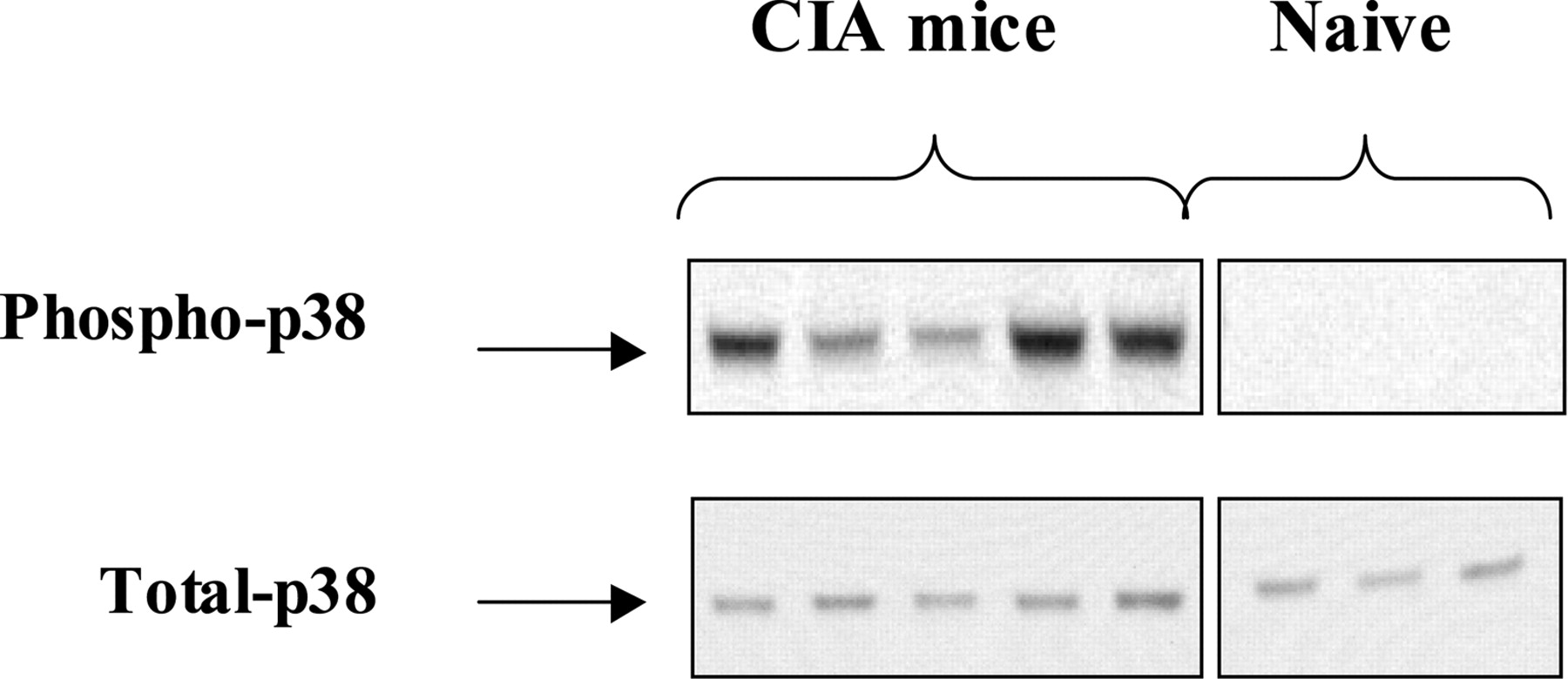
Phosphorylated p38 MAPK in paws from naive mice and mice with CIA: p38 MAPK is phosphorylated and activated in arthritic mice. Whole-mouse paw extracts (20 μg/lane) were subjected to SDS-PAGE and immunoblot analysis. Blots were probed with anti-phospho-p38 and anti-p38 MAPK antibodies. Immunodetection was visualized by enhanced chemiluminescence. Each lane represents inflamed paw lysate from a single animal. Results are representative of one experiment and a total of five arthritic and three naive mice.

Treatment with SD-282 improves clinical severity scores in mice with early CIA disease. Mice with early stage CIA were divided into three groups each of 15 mice. Groups were treated by oral gavage twice daily with vehicle (open circles) or 30 (open triangles) or 90 mg/kg SD-282 (filled circles) for 10 days. Values are reported as the mean ± S.D. *, p < 0.05; **, p < 0.001 versus vehicle, by nonparametric Bonferroni multiple comparison test. SD-282 reduced clinical severity scores in a dose-dependent manner.
Results
Collagen immunizations coupled with LPS treatment as a method to induce synovitis reminiscent of human arthritis was originally developed by Joosten et al. (1994, 1997). In our hands and as shown below, early signs of disease (slight swelling and erythema in one to two paws) are first noted 24 days following the first immunization with collagen. At the time of initiation of the study when the clinical scores are in between 1 and 2 (i.e., day 24), p38α MAPK is present in joint tissue in an active state as judged by Western analysis, but not in tissue from nondiseased control animals (Fig. 1). In general, it appears that mild phosphorylation of p38 levels correlates with a clinical score of 1 (Fig. 1, lanes 2 and 3), whereas moderate phosphorylation of p38 levels for paws correlates with clinical scores of 2 (Fig. 1, lanes 1, 4, and 5). Signs of the disease progressed, and by day 33, animals had pronounced swelling and erythema and joint rigidity in three to four paws. Histological assessment of mice at day 33 revealed significant pannus, joint space narrowing, and synovitis with IL-1-producing neutrophils and IL-6-producing monocyte macrophages present. In addition, osteoclasts were abundant, and bone erosion and cartilage loss were prominent features of joint lesions. In the studies reported here, treatment was initiated in animals with early (day 24) and advanced (day 33) arthritis.

Treatment with SD-282 inhibits IL-6, IL-1β, and COX-2 mRNA induction in arthritic paws of mice with early CIA disease. Taqman analysis of inflamed mouse paw tissues. Values are reported as the mean ± S.D. n = 6. Note the diminished mRNA expression of IL-6 (*, p < 0.04 versus vehicle by two-tailed Student's t test; A), IL-1β (*, p < 0.05 versus vehicle by two-tailed t test; B), and COX-2 (*, p < 0.005 versus vehicle by two-tailed t test; C).
SD-282 Attenuates CIA in Mice with Early Stage Disease. Initiation of vehicle treatment of mice with early stage of the disease (mean clinical severity scores of 1.6 ± 0.6 on day 24) resulted in a progressive worsening of signs of disease, achieving a maximal clinical severity score of 7 ± 3.3 by day 34. SD-282 treatment resulted in a dose-dependent improvement in clinical severity scores (Fig. 2) and paw swelling as measured by plethysmometer. Swelling, expressed as mean percentage increase of paw size compared with the baseline value, was 175 ± 100% for the vehicle-treated group, 90 ± 55% for animals treated with SD-282 at 30 mg/kg, and 47 ± 43% for animals treated with SD-282 at 90 mg/kg (p < 0.001 compared with the vehicle group). SD-282 at 90 mg/kg treatment was also associated with a reduction mRNA levels for proinflammatory genes including IL-1β, IL-6, and COX-2 in paw tissue (Fig. 3). Intriguingly, TNF-α mRNA levels are very low in arthritic mouse paw tissues in the vehicle group at the end of the study. This precluded our ability to accurately assess the effect of SD-282 on TNF-α at the end of the early stage treatment mode that falls on day 34 and can be considered as advanced stage disease for untreated animals. This suggests that TNF-α expression in the joint was not elevated in animals with advanced disease compared with the naive healthy mice. TNF-α mRNA levels were not significantly elevated at the time point used in this study, consistent with previous reports showing no clear peak of TNF mRNA expression in the mouse CIA model (Rioja et al., 2004).
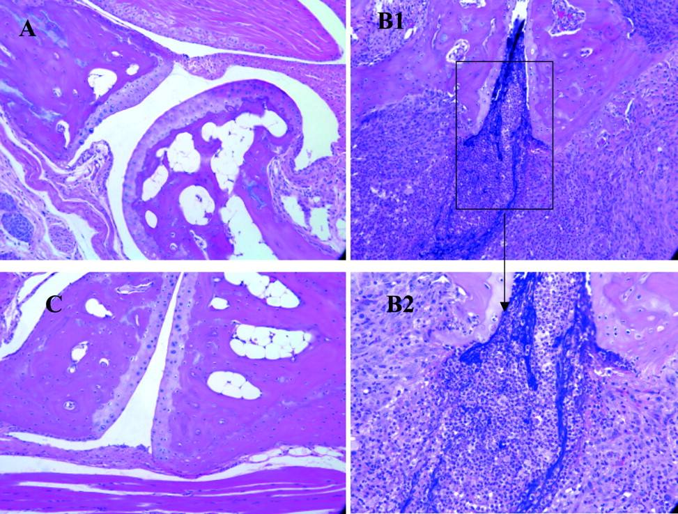
Treatment with SD-282 ameliorates cartilage and bone loss in mice with early CIA disease. Note normal bone joint in naive group (A), severe inflammation and cartilage and bone destruction in vehicle group (B1 and B2), and reduced inflammation and ameliorated cartilage and bone pathology in SD-282 at 90 mg/kg group (C).
Histological assessment of knee joints from vehicle-treated animals showed clear evidence of synovitis with proliferating pannus, bone erosion, cartilage destruction, and mononuclear cell infiltrate (Fig. 4). There was little evidence of any inflammation or other disease-related changes, including a loss in joint space in paw tissue from the SD-282 treatment group.
The effect of withdrawal of the p38α MAPK inhibitor on disease severity scores was evaluated in a second study. SD-282 treatment (30 and 90 mg/kg) when administered for 10 days from days 25 to 35 resulted in a dose-dependent improvement in clinical severity scores as seen in the first study. At the time of treatment withdrawal (i.e., day 35), clinical severity scores in vehicle, 30 mg/kg SD-282, and 90 mg/kg SD-282 groups were 9.7 ± 1.6, 4.2 ± 1.5, and 2.2 ± 1, respectively. Cessation of SD-282 treatment from day 35 onwards was not associated with any further increase of clinical scores when mice were monitored for an additional 10 days (i.e., up to day 44). On day 44, clinical severity scores in vehicle, 30 mg/kg SD-282, and 90 mg/kg SD-282 were 11 ± 1, 4.4 ± 1.6, and 2.2 ± 1.2, respectively. This is a durable effect, as evidenced by steady clinical severity scores for up to 44 days of evaluation.

Treatment with SD-282 improves clinical disease scores in mice with advanced CIA disease. Mice with advanced CIA (30 days after immunization) were divided into three groups each of 12 mice. Groups were treated by oral gavage twice daily with vehicle (open circles) or 30 mg/kg SD-282 (open triangles) or 90 mg/kg (hatched circles) for 20 days. Values are reported as the mean ± S.D. *, p < 0.05; **, p < 0.001 versus vehicle, calculated by nonparametric Bonferroni multiple comparison test. SD-282 significantly reduced clinical severity scores in a dose-dependent manner.
SD-282 Reverses Cartilage and Bone Destruction in Mice with Advanced Disease. In the first study, arthritic mice with advanced disease on day 30 were treated for 20 days with vehicle or SD-282 (30 and 90 mg/kg b.i.d.) by oral gavage. At the time of treatment initiation, clinical severity scores in the vehicle, 30 mg/kg SD-282, and 90 mg/kg SD-282 groups were 9.1 ± 2.6, 9.0 ± 3.2, and 9.1 ± 2.6, respectively. SD-282 treatment was associated with a dose-dependent, statistically significant improvement in clinical severity scores (Fig. 5), and paw swelling was measured by plethysmometer. At the end of the treatment period, clinical severity scores in the vehicle, 30 mg/kg SD-282, and 90 mg/kg SD-282 groups were 9.1 ± 2.2, 6.9 ± 1.6 (p < 0.05), and 4.9 ± 1.7 (p < 0.001)., respectively Paw swelling at day 50 (expressed as mean percentage increase in paw size compared with baseline) was 156 ± 16% in vehicle-treated animals, 142 ± 26% in animals treated with SD-282 at 30 mg/kg, and 120 ± 16% (p < 0.01 compared with vehicle group) in animals treated with SD-282 at 90 mg/kg. Cartilage and bone destruction were evaluated by histology and three-dimensional bone analysis. Histological assessment comparing tissue taken at the end of dosing from animals administered SD-282 or vehicle demonstrated a reversal in bone and cartilage destruction in the SD-282 treatment group (data not shown). The vehicle group demonstrated greater bone erosion and destruction (as assessed by three-dimensional bone analysis) than did SD-282 treatment groups (Fig. 6, A and B), consistent with increased bone resorption and heterotopic ossifications. Bone destruction was reversed as evidenced by the fact that bone volume fraction and thickness were statistically significantly greater in the SD-282-treated groups compared with the vehicle group (Table 2). The three-dimensional images show greater bone erosions and destruction because of increased bone resorption and heterotopic ossifications in vehicle-treated mice than in SD-282-treated mice.
SD-282 reverses bone destruction as evidenced by bone volume, thickness, and separation

Three-dimensional μCT reconstructions of the rear paws showing that SD-282 reverses bone erosion. The cortical bone and articular surface of the metatarsals I to V, the cuneiforms I to III, the cuboid, the navicular, the calcaneus, the talus, the distal fibula, and the distal tibia can be observed in these images. There are severe erosions in all of the bones in the right paw from the vehicle group (A). No erosion can be observed in the paw from the group receiving treatment with 90 mg/kg SD-282 (B).
To confirm the results of a prior study and to collect histological and bone data at the time of treatment initiation, a second study was performed. In this follow-up study, mice with signs of advanced disease (severity scores of 9.6 ± 1.8, day 33) were treated orally with vehicle or SD-282 at 90 mg/kg for 28 days, i.e., up to day 61. In the vehicle treatment group, clinical severity scores remained high through the end of the study (9.6 ± 1.8 and 9.3 ± 0.9 at the beginning and end of the treatment period, respectively). SD-282 treatment improved clinical severity scores in a statistically significant manner (p < 0.0003) from a baseline of 9.5 ± 1.8 at the beginning of the study (day 33) to 5.5 ± 2 by the end of the study (day 61). SD-282 at 90 mg/kg also reduced paw swelling as measured by plethysmometer (data not shown). After 28 days of treatment, body weight gain was higher in the SD-282 group compared with the vehicle group (4 ± 1 and 2.8 ± 0.7 g, respectively; p < 0.01).
In both experiments, cartilage and bone destruction was evaluated by histology. These two studies generated similar data on cartilage and bone destruction. Therefore, data in the second study are reported in this communication. To confirm that SD-282 has a key role in cartilage and bone destruction in joint disease, we graded pathology on sections of whole knee joints. In the naive group, no abnormal histopathological changes were observed (Fig. 7A). Histological examination of paw tissue from the baseline group revealed arthritic lesions clearly evident in both front and hind paws and in both small and large joints (Fig. 7B). Histopathological changes including cartilage erosion, bone erosion, joint destruction, synovitis, pannus formation, and synovial fibroplasia were observed in all of the examined paws. In the vehicle-treated group at the end of the experiment (i.e., on day 61), the development of arthritis was obvious in both front and hind paws and in both small and large joints (Fig. 7C). Histopathological changes were observed in every one of the vehicle-treated paws In contrast, for the SD-282-treated group, no markedly severe arthritis was observed in any of the paws (Fig. 7D). Each of the six histology parameters (cartilage, bone erosion, joint destruction, synovitis, pannus, and fibroplasias) was lower in a statistically significant manner (Fig. 8). In the vehicle-treated group, osteoclasts (large multinuclear cells implicated in pathologic bone loss) were located at the site of bone destruction (arrows, Fig. 9). Treatment with SD-282 significantly decreased the number of osteoclasts (1.5 ± 1.5) compared with the vehicle (6 ± 2) at day 61 (p < 0.03).
Immunohistochemistry of COX-2, IL-1β, and IL-6. In the naive group (no induction of arthritis and no treatment), COX-2, IL-1β, and IL-6 were not observed in any sample by the staining methods used here. In the baseline group (representing time of treatment initiation after induction of arthritis), IL-1β staining was found in inflammatory cells (primarily neutrophils) within the joint area. IL-1β staining was significantly lower in the SD-282 group compared with the vehicle group (p < 0.02; Fig. 10, A1, B1, and C1; Table 3). IL-6 staining, primarily observed in macrophages, was low at baseline and was greater in the vehicle group than in the SD-282 group (p < 0.007; Fig. 10, A2, B2, and C2; Table 3). In the baseline group, COX-2 was detected in chondrocytes and synovial fibroblasts. At the end of the treatment period, COX-2 staining was significantly reduced in the SD-282 treatment group compared with the vehicle group (p < 0.01; Fig. 10, A3, B3, and C3; Table 3).
SD-282 lowers COX-2, IL-β, and Il-6 expression in the joint tissue of CIA mice with advanced disease

SD-282 reverses cartilage and bone loss and promotes bone healing in mice with advanced CIA. SD-282 treatment improves histological scoring. Mice showing signs of advanced disease on day 33 were randomized to three groups: 1, baseline animals, sacrificed on day 33; 2, 28-day vehicle treatment, sacrificed on day 61; and 3, 28-day treatment with SD-282 at 90 mg/kg, sacrificed on day 61. Knee joint histology from representative naive mice (A), baseline on day 33 (B), and treatment at day 61 with vehicle (C) and SD-282 at 90 mg/kg (D) are shown. Knee joint histology of vehicle-treated mice on day 61 reveals severe bone and cartilage destruction, pannus accumulation, neutrophil, and macrophage (ED-1 staining) infiltration. Histology from the SD-282 treatment group at day 61 (D) reveals marked improvement in joint histology including evidence of cartilage and bone healing.

SD-282 reverses cartilage, bone, and joint destruction as well as synovitis, pannus, and fibroplasia in mice with advanced CIA. Mice showing signs of advanced disease were selected 33 days after initial collagen immunization and divided into three groups. One group (base-line) was sacrificed on day 33. The other two groups were treated with vehicle or SD-282 at 90 mg/kg daily until day 61. Open bars, histopathology scores of paws from baseline, i.e., day 33; hatched black bars, scores of paws from the vehicle-treated group at day 61; hatched gray bars, scores of paws from SD-282 at 90 mg/kg treated group at day 61. SD-282 significantly reduces cartilage erosion (**, p < 0.01), bone erosion (*, p < 0.05), joint destruction (***, p < 0.001), synovitis (**, p < 0.01), pannus (***, p < 0.0001), and fibroplasia (**, p < 0.01) compared with the vehicle-treated group. Values are reported as the mean ± S.D. Statistics were calculated by nonparametric Bonferroni multiple comparison test.
SD-282 Reverses Bone Erosion in Advanced CIA as Evidenced by Three-Dimensional μCT Analysis. Three-dimensional μCT studies revealed a complete lack of bone erosion in samples from the naive group. In contrast, bone erosion was significantly evident in the animals with CIA at baseline on day 33. The erosion was more pronounced in the vehicle group on day 61 (erosion score, 32 ± 15) than at baseline (erosion score, 12.7 ± 4.6). The erosion score of the SD-282-treated group at day 61 was 3.5 ± 2.9, apparently reversing erosion present at baseline, restoring the bone back to almost naive levels (p < 0.0001 compared with the vehicle group). Three-dimensional μCT bone erosion scores show that the score in the vehicle group is approximately 2.5-fold over baseline, whereas the score in the SD-282 treatment group is over 9-fold lower than the vehicle group and approximately 3.5-fold lower than at baseline. Three-dimensional morphometric measurement shows that SD-282 treatment significantly reverses loss in the bone volume fraction and bone microarchitecture (data not shown).
COMP, A Circulating Marker of Cartilage Turnover, Is Decreased by SD-282. To obtain further insight into the protection against cartilage destruction, we determined serum COMP levels in the naive, vehicle, and SD-282 groups of the second experiment. COMP is released from cartilage as a result of increased turnover in human and experimental arthritis. Circulating COMP levels were 2.1 ± 0.4, 6.7 ± 1, and 5.2 ± 0.7 in naive, vehicle, and SD-282 at 90 mg/kg groups, respectively. SD-282 significantly reduced COMP levels compared with the vehicle group (p < 0.001).
Discussion
Although the relative expression of p38 MAPK isoforms in rheumatoid arthritis has not been fully explored, p38α and p38δ MAPKs are known to be especially prevalent at sites of joint destruction (McLay et al., 2001). Here, we show that at day 24, an early phase of the disease, p38α MAPK, is in an active (phosphorylated) state in tissue from arthritic joints compared with nonarthritic tissue. Although we did not evaluate the phosphorylation state of p38α MAPK at time points later in the disease process, it is clear from histology that synovitis is present at days 34 and 61 in animals with symptoms, suggesting that p38α MAPK remains active in tissue from later disease stages. SD-282 demonstrated selectivity for the α isoform, showing 14.3 to >1000 times greater potency against p38α MAPK than it did against p38β-, p38γ-, and p38δ-MAPKs. To further understand the role of the α isoform in arthritic disease and to explore the therapeutic potential of SD-282 on bone and cartilage destruction, we proceeded to test this α-specific inhibitor in mice with CIA disease. In particular, we were curious to see whether SD-282 would have beneficial effects in late stage disease, when bone and cartilage destruction become manifest.

SD-282 reduces osteoclast number in mice with advanced CIA. Note arrows in A1 and A2 of vehicle-treated group for abundant osteoclasts. SD-282 treatment (B1 and B2) almost removed those osteoclasts in joint tissue from CIA mice with advanced disease on day 61.

SD-282 reduces IL-1β, IL-6, and COX-2 expression in inflamed joints in mice with advanced CIA. Mice with joint histology showing signs of advanced disease on day 33 were randomized to three groups: column A, baseline animals, sacrificed on day 33; column B, 28-day vehicle treatment, sacrificed on day 61; and column C, 28-day treatment with 90 mg/kg SD-282, sacrificed on day 61. Sections were probed for expression of IL-1β (row 1), IL-6 (row 2), and COX-2 (row 3) in the pannus of synovium and in the chondrocytes of cartilage by immunohistochemistry. SD-282 treatment at day 61 was associated with marked reduction of IL-1β (C1), IL-6 (C2), and COX-2 (C3) expression compared with the corresponding vehicle groups (B1, B2, and B3) as well as baseline groups (A1, A2, and A3).
It has been reported that a nonspecific p38 MAPK inhibitor attenuates arthritis in the early stage of the mouse CIA disease (Nishikawa et al., 2003). As expected from the anti-inflammatory profile of p38α MAPK inhibitors and previous studies in animals with experimental arthritis (Badger et al., 1996; Kumar et al., 2003; Nishikawa et al., 2003), SD-282 treatment of animals with early stage arthritis reduced the progression of the disease. This effect was evidenced by improvements in clinical severity scores, histological assessments (pannus, synovitis, cartilage, and bone destruction), and reduced inflammatory gene expression (IL-1β, IL-6, COX-2). Furthermore, this study also revealed that arthritis symptoms did not recur upon withdrawal of SD-282 treatment from mice treated at an early stage of disease.
Although several interventions have shown utility in early stage CIA in mice and in human RA, control of bone and cartilage destruction of the sort seen in advanced CIA in mice is the most challenging objective in the treatment of human RA. Several lines of evidence demonstrate that murine CIA progresses to an advanced state that is comparable with that seen in humans. In areas of tumor-like synovial tissue, erosion of trabecular and cortical bone is common, leading to the characteristic erosions seen on radiography. In our model, osteoclasts can be seen in the areas of bone destruction during CIA. Furthermore, in patients with rheumatoid arthritis, elevated serum COMP levels are an important marker of cartilage degradation, and treatment with TNF-α inhibitors (infliximab or etanercept) has been shown to reduce COMP levels (Larsson et al., 1997; Joosten et al., 1999b; Crnkic et al., 2003). We detected elevated COMP levels in vehicle-treated over naive mice.
Here, we report for the first time an oral, α-specific inhibitor of p38 MAPK effective in the treatment of advanced CIA in mice. SD-282 treatment of mice with advanced arthritis resulted in significant improvement in clinical scoring and a decrease in paw swelling compared with treatment with vehicle alone. Histological assessments comparing SD-282 and vehicle treatment cohorts revealed statistically significant results consistent with an interpretation of striking reversal in all aspects of joint disease, including reduced osteoclast number, synovitis, pannus, fibroplasia, an increase in joint space, and decreased erosions in bone and cartilage (Figs. 7, 8, 9). The interpretation of positive reversal effects of SD-282 treatment on bone was also supported by improved bone parameters measured by μCT, including bone volume index, structural thickness, and structural separation (Fig. 6, A and B; Table 2). In addition, the improvement in cartilage in the SD-282 treatment group was accompanied by significant reduction in COMP levels.
It is well established that p38α MAPK regulates key proinflammatory molecules in RA, including TNF-α, IL-1β, IL-6, and COX-2. In the present study, we have shown that in animals with advanced disease, IL-1β, IL-6, and COX-2 are prominent in the joint lesion, and their expression is reduced with p38α MAPK inhibitor treatment. Using the same mouse arthritis model employed here, Joosten et al. (1996) demonstrated that anti-TNF-α treatment had no beneficial effect in animals with advanced disease, suggesting that this cytokine is less important in advanced stage of the disease in this model. This is consistent with our observation that TNF-α expression in the joint was not elevated in animals with advanced disease compared with naive healthy mice. It is generally believed that TNF-α is important in early stages of this disease model (Williams et al., 1992; Wooley et al., 1993) but not in advanced disease (Joosten et al., 1996). IL-6 has been shown to be associated bone resorption (Palmqvist et al., 2002); more precisely, osteoclastogenesis (Li et al., 2004) and its inhibition reduces bone resorption (Suzuki et al., 1998; Nishikawa et al., 2003). IL-1β blockade prevents cartilage and bone destruction in mouse CIA arthritis, whereas TNF-α blockade only ameliorates joint inflammation (Joosten et al., 1999a). Inhibition of COX-2 has been associated with reduced cartilage and bone destruction in rat adjuvant-induced arthritis, suggesting that this enzyme is important for regulating disease progression (Chan et al., 1999). Thus, effects on IL-6, IL-1β, and COX-2 may alter the balance between osteolysis and osteogenesis in animals with advanced experimental arthritis.
Interestingly, these studies demonstrate that in a model of advanced arthritis associated with significant osteolysis, inhibition of p38α MAPK correlates with statistically significant reversal of cartilage and bone destruction. A comparison of histology from arthritic animals in the baseline group and the SD-282 treatment group shows a striking improvement in bone density and cartilage integrity, suggesting not only a reversal but also a bone-healing effect. Bone healing is also evidenced by significantly improved quantitative bone erosion scores between the baseline and SD-282 drug treatment groups. Erosion scores in the vehicle treatment group were even greater than those from the baseline group, suggesting that bone erosion progresses in the absence of pharmacological intervention. The histological improvement in cartilage seen in the SD-282 group compared with baseline suggests that, in addition to reversing the disease, SD-282 also has a positive effect on bone and cartilage healing.
It appears that this bone healing could be achieved through a mechanism involving both bone resorption/osteoclastogenesis and bone formation/osteoblastogenesis. Available literature clearly indicate that p38 MAPK is a potent mediator of osteoclastogenesis and osteoclast differentiation mediated by a wide variety of inducers including TNF-α, receptor activator of nuclear factor-κB ligand, and PGE2 (Nishikawa et al., 2003; Steeve et al., 2004). p38 MAPK inhibition has been shown previously to block bone resorption in fetal rat long bones in vitro (Kumar et al., 2001). In the study reported here, inhibition of p38α MAPK was associated with reduced osteoclasts. It is doubtful; however, that this was sufficient to explain the extent of bone healing seen. Along with osteoclasts, other cell types (synovial fibroblasts and macrophages) produce enzymes and factors that could contribute to bone erosion. In mice with advanced arthritis, we noted reduced synovitis and reduced osteoclast numbers in the vehicle treatment group compared with baseline; however, joint space narrowing and bone and cartilage destruction was not reduced. In fact, by quantitative μCT, bone erosion scores continued to progress in the vehicle-treated group but not in the SD-282-treated group. Indeed, by day 61, erosion scores for the SD-282 treatment group achieved a level between those seen in the naive (nonarthritic mice) and the baseline (start of therapy) group. This suggests that SD-282 healed bones in the late stage of CIA disease by reducing bone resorption and by forming new bone. We hypothesize that the effect of SD-282 on bone may be due to inhibition of osteoclast differentiation in the absence of IL-6 (Li et al., 2002, 2003; Yamamoto et al., 2003) and/or osteoblastogenesis. We are in the process of testing the latter hypothesis. The effects on the cartilage may be due to inhibition of signaling cascades that mediate the up-regulation of COX-2 expression and PGE2 production in chondrocytes exposed to the proinflammatory cytokine IL-1β and/or elevation of Col2A1 gene expression in the absence of IL-1β (Goldring and Berenbaum, 1999; Nieminen et al., 2005). The loss of pannus tissue is a striking feature of SD-282 treatment. The exact mechanism resulting in this effect is not known. However, proinflammatory pathways are believed to promote the expansion and maintenance of pannus tissue (Desmoulins et al., 1990; Paleolog et al., 1998). In this regard, the anti-inflammatory actions of p38 MAPK inhibition probably contribute to the effect on pannus noted in these studies. Further studies on the role of SD-282 in new bone formation with an aid of histomorphometry are warranted.
The present study indicates that inhibiting p38α MAPK during advanced stages of murine collagen-induced arthritis improves all aspects of the disease including synovitis, joint space narrowing, and bone and cartilage structures. The inhibition of osteolytic lesions and reversal of cartilage and bone destruction reported in this model reflects the modulation of multiple p38α MAPK-dependent pathways including IL-1β, IL-6, and COX-2. Thus, these studies suggest that inhibition of p38α MAPK has the potential to offer a unique therapeutic strategy for treating advanced stages of RA.
Footnotes
-
Article, publication date, and citation information can be found at http://jpet.aspetjournals.org.
-
doi:10.1124/jpet.105.098020.
-
ABBREVIATIONS: RA, rheumatoid arthritis; TNF, tumor necrosis factor; IL, interleukin; COX, cyclooxygenase; MAPK, mitogen-activated protein kinase; SD-282, indole-5-carboxamide (ATP-competitive inhibitor of p38 kinase), CIA, collagen-induced arthritis; PGE2, prostaglandin E2; FR167653, pyridinyl imidazole compound (specific inhibitor of p38 pathway); LPS, lipopolysaccharide; ELISA, enzyme-linked immunosorbent assay; SB202190, (4-(4-fluorophenyl)-2-(4-hydroxyphenyl)-5-(4-pyridyl)1H-imidazole; COMP, cartilage oligomeric matrix protein; 3-D, three-dimensional; micro-CT, μCT, microcomputed tomography.
- Received November 2, 2005.
- Accepted March 24, 2006.
- The American Society for Pharmacology and Experimental Therapeutics


